Quantcast
Channel: Fun With Microbiology (What's Buggin' You?)

Apophysomyces elegans "complex"

$
0
0


Apophysomyces elegans complex(Zygomycetes) Order Mucorales

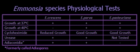
Note:As of this blog post, most textbooks still list Apophysomyces elegans as the sole species in this genus.  Recent studies [i]have demonstrated a high variability among the 5.8S rRNA gene sequences of clinical strains of A.elegans.  This study suggests that A.elegans is actually a complex composed of several (three) newly proposed species.  They have proposed the species names A.ossiformis, characterized by bone-shaped sporangiospores, A.trapeziformis, with trapezoid-shaped sporangiospores, and A.variabilis with variable-shaped sporangiospores.  A.elegans remains as the fourth species of the Apophysomyces complex.  Physiologically, A.elegans is able to assimilate the glycoside esculin, whereas the three newly proposed species failed to assimilate esculin.  Recent studies suggest that A.elegans may actually be a non-pathogenic environmental species, while the remaining three species are etiologically linked to clinical disease.  Further studies are warranted.

Ecology:
Although the ecology of Apophysomyces is rather poorly described, this fungus appears to have widespread distribution and has been isolated from soils and decaying vegetation in India, Australia, Southeast Asia and the United States.

Pathology:
Apophysomyces is an occasional agent of Zygomycosis.  Infection by Apophysomyces species differs from other Zygomycetes infections in two ways.  Firstly, it occurs more frequently in immunocompetent than immunocompromised individuals, whereas other Zygomycetes primarily infect hosts with weakened immune systems.  Secondly, Apophysomyces infections are acquired directly by traumatic implantation rather than by inhalation of spores which then progress to rhino-cerebral or other disseminated infections.

Apophysomycesinfection may result in necrotizing cellulitis or fasciitis (flesh eating) requiring aggressive surgical debridement and antifungal therapy.  Despite immediate and aggressive intervention, the prognosis is often poor as once established the fungus can quickly spread to adjacent tissues and distant sites via the blood stream. 

In 2011, a tornado carved a path of destruction through the American town of Joplin Missouri killing 160 citizens.  Many others were received traumatic injuries and of those, there were there were thirteen cases of necrotizing cutaneous zygomycosis due to Apophysomyces trapeziformis, five of which were fatal.

While the descriptions of Apophysomyces in this blog is gathered from a variety of sources under the name of Apophysomyces elegans, the species presented photographically in this blog post is Apophysomyces variabilis.

Macroscopic Morphology:
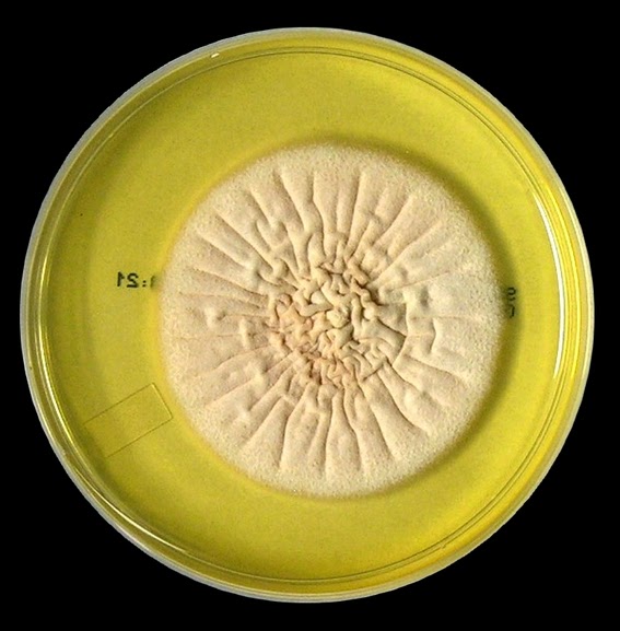
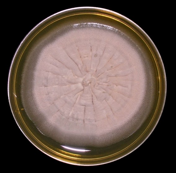
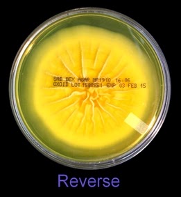
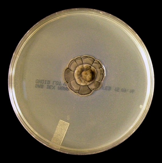
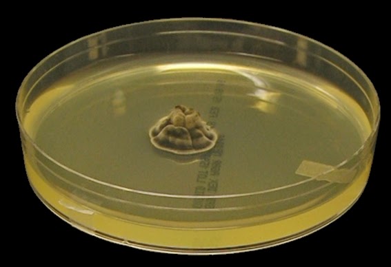
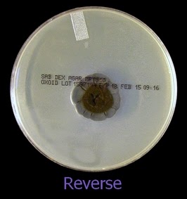
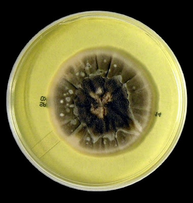
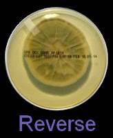
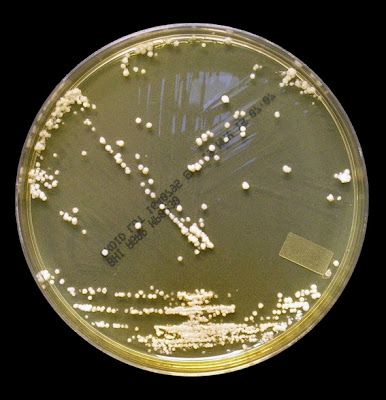
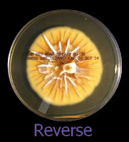
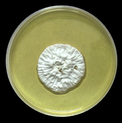
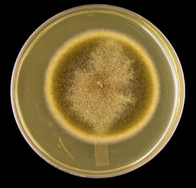
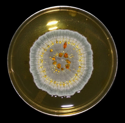
As with other Zygomycetes, Apophysomyces exhibits very rapid growth, often filling the petrie dish with profusely woolly mycelia in two to three days.  Growth (SAB, 30ᵒC) initially appears white or off-white in colour but may acquire a slight brownish-grey colour as the colony ages.  The reverse is white to pale yellow.

 Apophomyces variabilis - on SAB, 48 Hours at 30ᵒC (Nikon)

 Apophomyces variabilis - on SAB, 48 Hours at 30ᵒC (Nikon)

Microscopic Morphology:
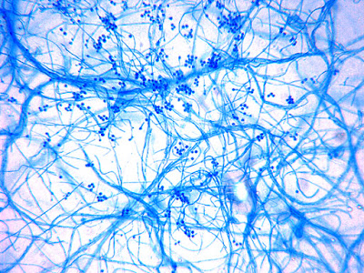
Note: The isolate presented here arrived in our laboratory as a proficiency testing challenge.  An initial direct tease mount revealed very broad, aseptate hyphae, suggesting Zygomycetes.  The isolate grew exceedingly well (SAB, 30ᵒC) yet failed to produce any fruiting structures.  The combined observations of broad aseptate hyphae and failure to produce fruiting structures on routine mycological media raised suspicions that this isolate belonged to either an Apophysomycesspecies, or was Sakseneavasiformis.

Both Apophysomyces species and Saksenea vasiformisare known to be notoriously resistant to efforts attempting to induce sporulation.  Study of the structures associated with sporulation greatly assists the identification of the Zygomycetes and as such, techniques have been developed in an attempt to induce the spore production[ii].   Unfortunately, our laboratory supports an acute care hospital and we lack the facilities for any experimentation outside of the routine clinical protocol.  (ie. purchased prepared media: no autoclave, media components, etc.)  However this blog is entitled “Fun With Microbiology” and I had “fun” improvising the ‘sterile water and yeast extract sporulation media’ outlined in endnote ii with some success.

Microscopic Morphology Continued:
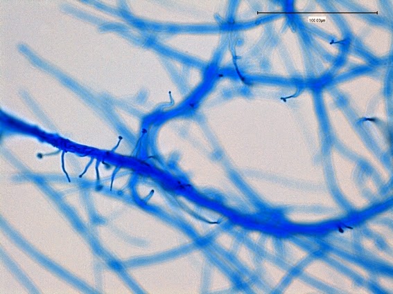
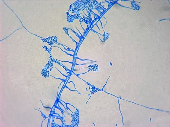
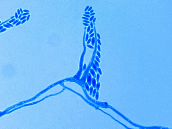
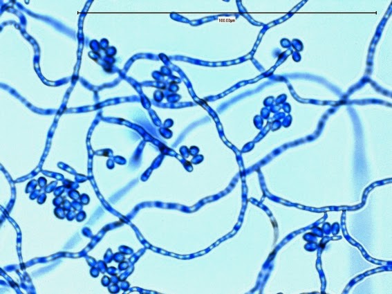
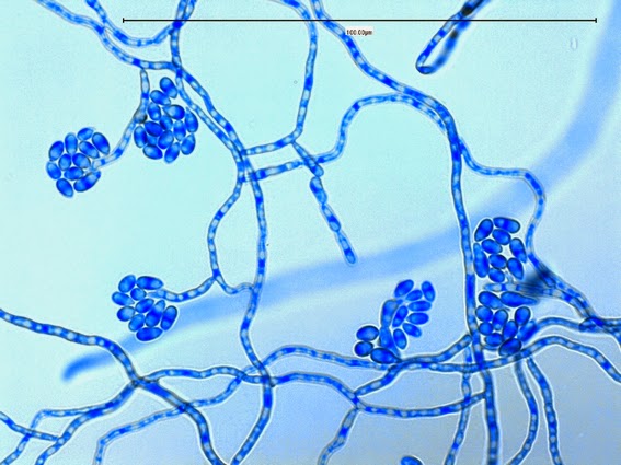
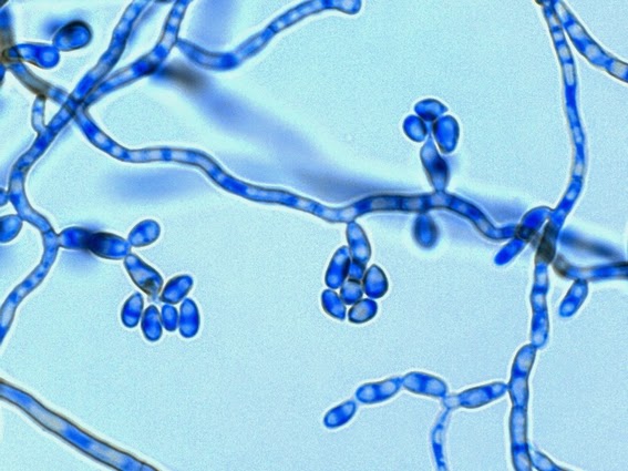
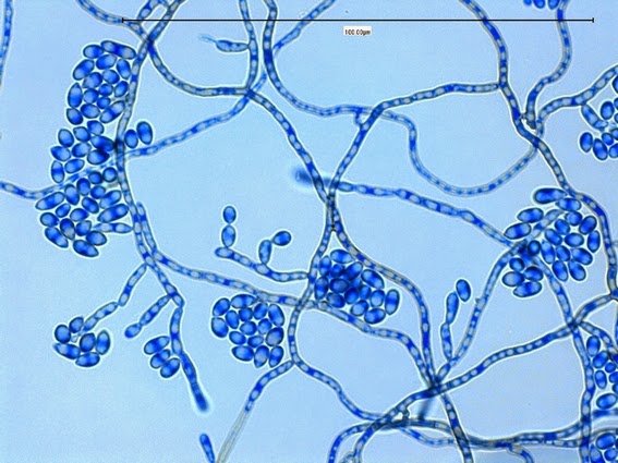
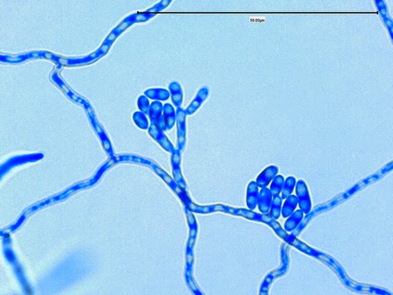
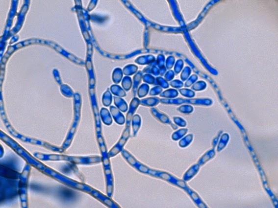
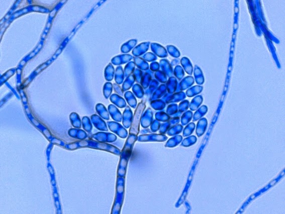
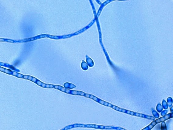
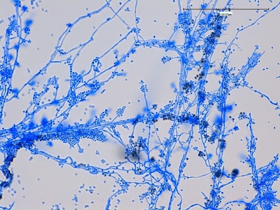
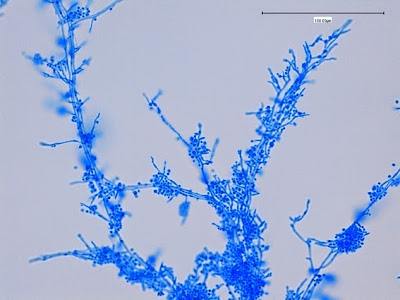
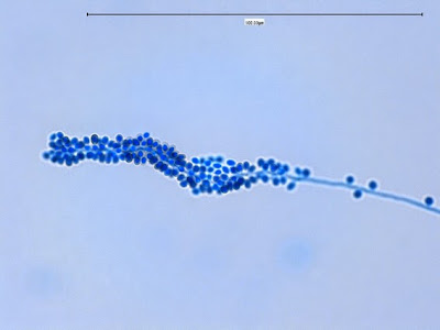
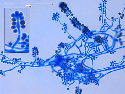
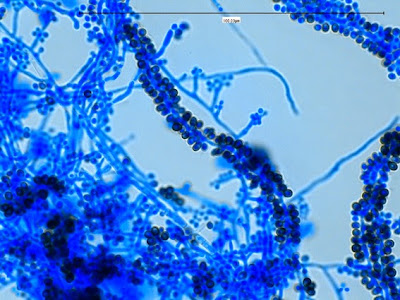
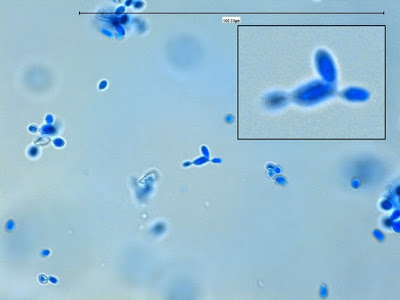
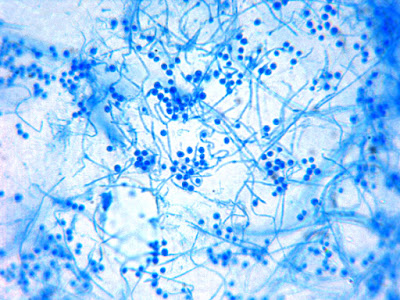
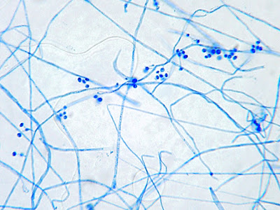
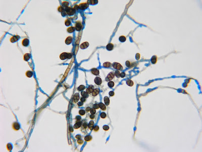
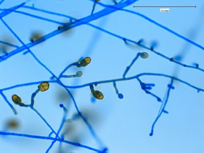
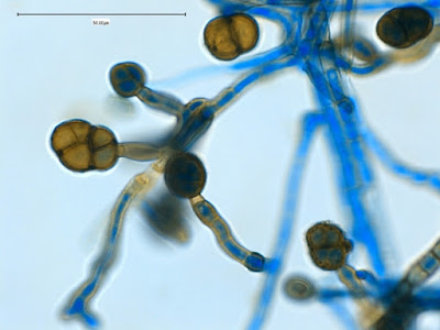
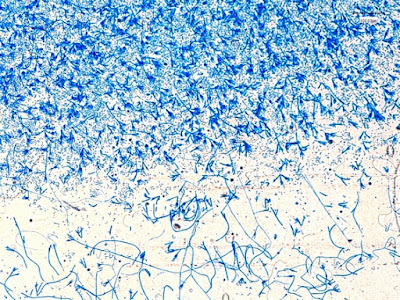
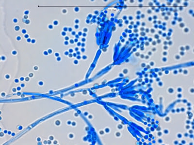
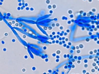
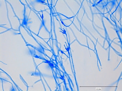
Apophysomyces species produce broad based (up to 10+ µm dia.) hyaline (clear, non-pigmented) hyphae which are almost entirely aseptate.  Sporangiophores are quite long (up to 540 µm), unbranched and usually produced singly from aerial hyphae.  Sporangiophores may be attached to the hyphae with a prominent structure resembling the ‘foot cell’ found in Aspergillus species.  The apex of the sporangiophore widens to form a structure called an apophysis (hence the genus name) which may be distinctly bell-shaped or vase-shaped (20 -58 µm dia.) distinguishing it from that of other Zygomycetes.  Yet other sources describe the apophysis as champagne glass shaped or perhaps more common to other Zygomycetes, funnel-shaped.  The columella (18 – 28 µm) is hemispherical (half of a sphere) in shape.  Sporangiospores (5.4 – 8.0 X 4.0 – 5.7 µm) have been described as smooth and subspherical to cylindrical in shape.  Recall from the initial ‘Note’ at the start of this blog, the newly proposed species are molecularly distinct but also appear to produce distinctly shaped sporangiospores.  Rhizoids are produced and are generally located beneath or to the side of the sporangiophores.

An apology: as I lacked the facilities to properly induce sporulation in this fungus, these photos are as good as I'm going to get.

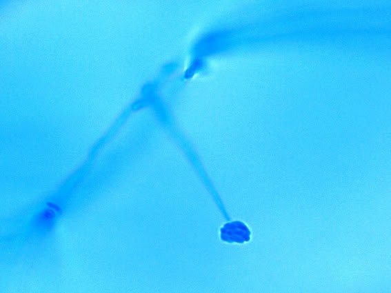
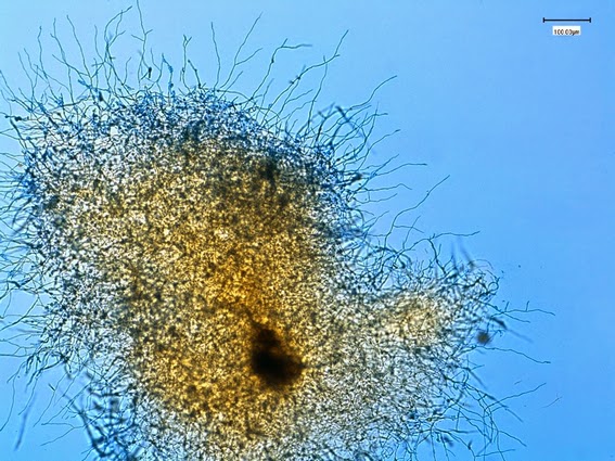
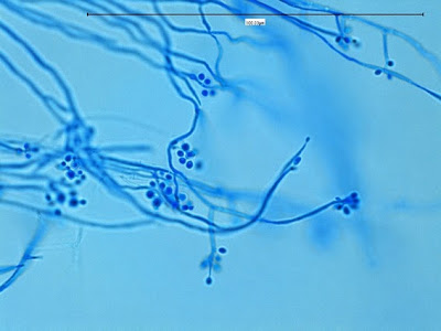
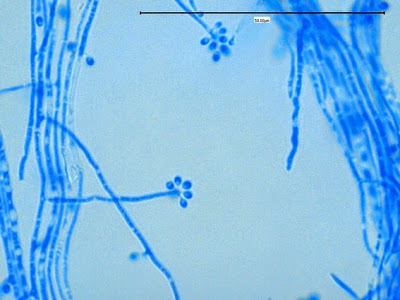
 Apophomyces variabilis - edge of tease mount showing single, well defined sporangiophore with attached sporangium.(250X, LPCB, Nikon)

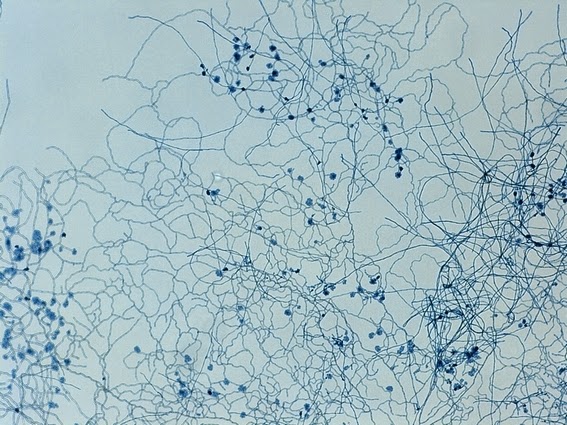
 Apophomyces variabilis -direct tease moount.
(400X, LPCB, Nikon)

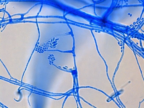
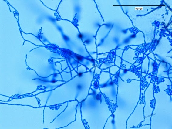
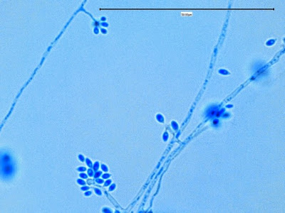
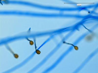
Apophomyces variabilis - apophysis without sporangium visible.  Several sporangiospores still visible in the bottom of the rather funnel-shaped apophysis.
(400X, LPCB, Nikon)

Apophomyces variabilis -again, from the direct tease mount showing a rather damaged apophysis remaining at the apex of the sporangiophore.
(400X, LPCB, Nikon)

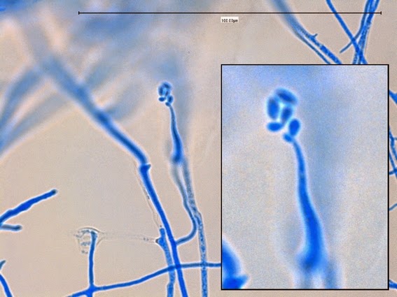
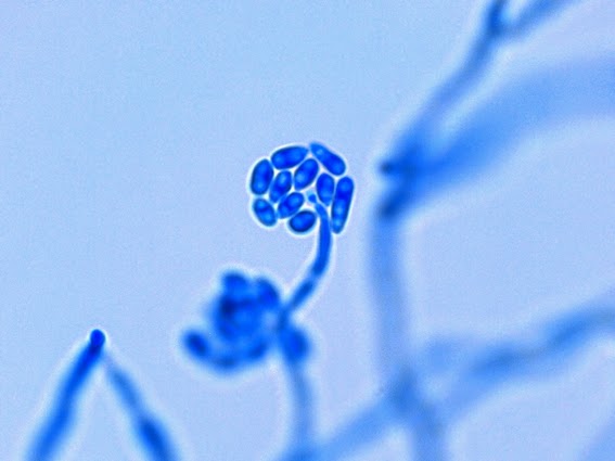
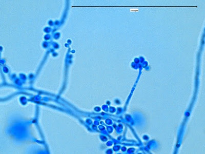
Apophomyces variabilis - young sporangiophore at the tip of the sporangiophore.
(400+10X, LPCB, DMD-108)

Apophomyces variabilis -as above but appears to be some development of the sporangiospores within the sporangium.  (400+10X, LPCB, DMD-108)

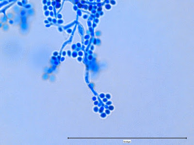
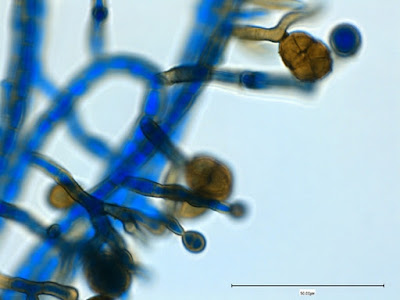
Apophomyces variabilis -maturing sporangium with sporangiospores visible within.
(400+10X, LPCB, DMD-108)

Apophomyces variabilis -much the same.
(400+10X, LPCB, DMD-108)

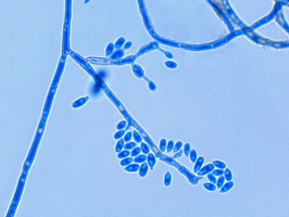
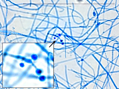
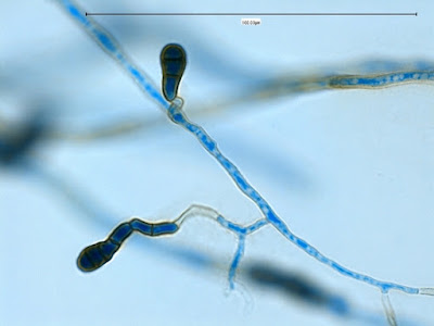
Apophomyces variabilis - produces long, un-branched sporangiophores.  One unique characteristic described in Apophysomyces elegans is a dark area (arrow) often found on the sporangiophore slightly below the apophysis.  (400+10X, LPCB, DMD-108)

Apophomyces variabilis -two sporangiophores.  The one on the bottom might be described as champagne flute-like in shape and there is at least one sporangiospore present within.
(400+10X, LPCB, DMD-108)

Apophomyces variabilis -probably an immature sporangiospore.
(1000X, LPCB, DMD-108)

Apophomyces variabilis -two columellas (?) with the one at the top of the photo showing sporangiospores still clinging to the surface.  The sporangium appears to have

dehisced (dissolved, split open).  (1000+10X, LPCB, DMD-108)

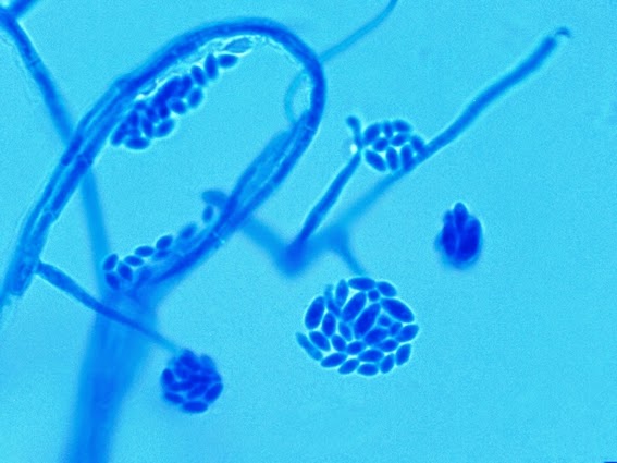
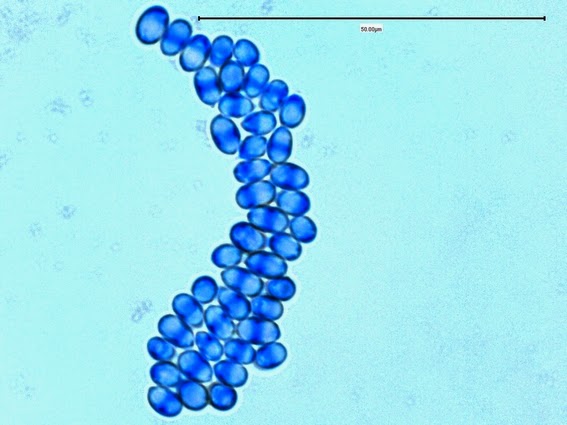
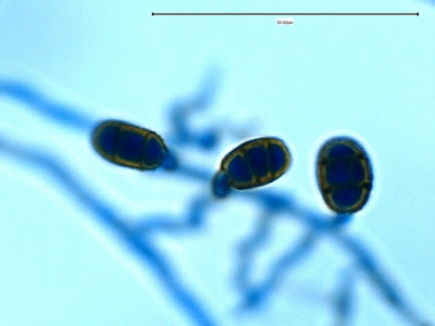
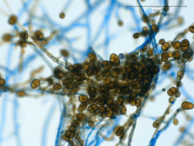
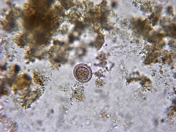
Apophomyces variabilis -looking down on a sporagium with a number of sporangiospores still attached to the surface.  Average size of the sporangiospores along the longer axis averaged 5.22 µm.
(1000+10X, LPCB, DMD-108) 

Apophomyces variabilis -an immature sporangium.
(1000+10X, LPCB, DMD-108)

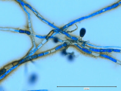
Apophomyces variabilis -more of the same.  The large width of the zygomycete hyphae is evident here as a hypha runs through the photo.  (1000+10X, LPCB, DMD-108)

Apophomyces variabilis -again, the broad hyphae of zygomyces in general.  The dimension reads 10.74 µm.  (400X, LPCB, DMD-108)

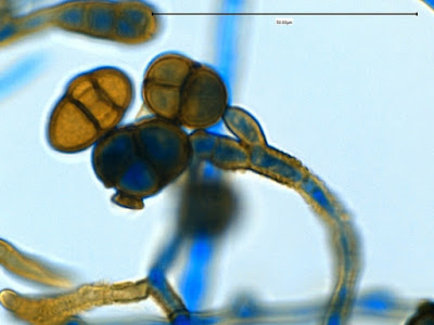
Apophomyces variabilis -the apophysis looks distinctly bell-shaped in this microphotograph.  Sporangiospores appear to be present within the developing sporangium.
(400+10X, LPCB, DMD-108)

Apophomyces variabilis -to try to convince you of the bell-shaped apophysis.  Rather than a funnel or cone shape where each wall runs on one continuous angle, the walls here start on one angle, the abruptly flare out at a greater angle, somwhat resembling a bell.
(400X, LPCB, DMD-108)

Apophomyces variabilis -a rather shorter sporangiophore with a rhizoid seen at the top of the photo and apophysis and broken sporangium spewing sporangiospores at the bottom of the photo.
(400X, LPCB, DMD-108)

Apophomyces variabilis -dissolved or broken sporangium seen at center-right of photo, again releasing sporangiospores.  Large bubble at lower left of picture spoils this photo.
(400+10X, LPCB, DMD-108)

Apophomyces variabilis -champagne flute-like apophysis with few sporangiospores still clinging to the interior.  (1000+10X, LPCB, DMD-108)

Apophomyces variabilis -sporangium is missing with the apophysis remaining at the apex of the sporangiophore.  Not at the base of the sporangiophore (right side), the foot-cell is clearly evident and has picked up an intense blue colour from the LPCB.
(400+10X, LPCB, DMD-108)

Apophomyces variabilis -foot-cell of Apophysomyces which resembles the same feature more commonly found in Aspergillus species.
(1000X, LPCB, DMD-108)

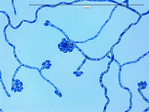
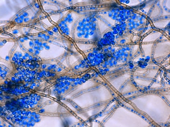
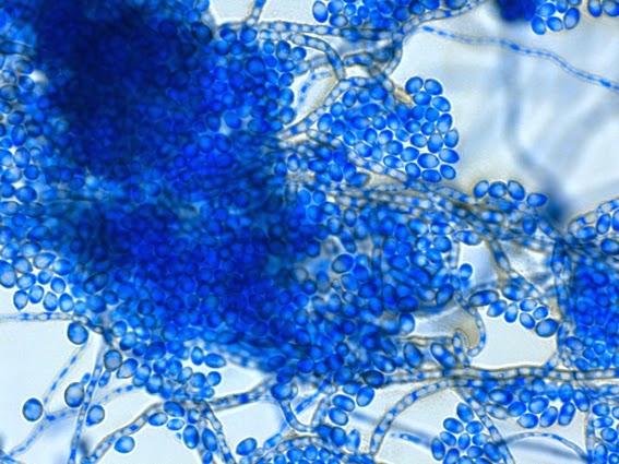
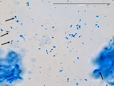
Apophomyces variabilis -a sporangium breaking apart to release the sporangiospores within.  They really do look variable in shape in this photo, true to the species name, 'variabilis'.
(1000+10X, LPCB, DMD-108)

Apophomyces variabilis -rhizoid, appears to have wrapped itself around a hyphal element.
(400X, LPCB, DMD-108)

Apophomyces variabilis -small rhizoid at center.
(400X, LPCB, DMD-108)

Apophomyces variabilis -okay, now that's a rhizoid! 
(1000X, LPCB, DMD-108)

Zygomycetes - some of the structures mentioned here and common to most zygomycetes


Physiology:
Apophysomyces, unlike other Zygomycetes, exhibits resistance to cycloheximide and therefore should grow on Mycosel® and Dermasel ® media.

Apophysomycesgrows well at 30ᵒC, 37ᵒC & 42ᵒC


Eduardo Alvearez, Alberto M. Stchigel, Josep Cano, Deanna A. Sutton, Annette W. Fothergill, Jagdish Chander, Valentina Salas, Michael G. Rinaldi and Josep Guarro
Rev. Iberoam Microbiol., 2010 27(2) pg. 80 - 89 (for purchase)
Arvind A. Padhye and Libero Ajello
Journ. Clin. Microbio., Sept. 1988, pg. 1861 – 1863.   (free PDF)                                                                 

Sepedonium species

$
0
0


Sepedonium species

Ecology:
Sepedoniumspecies are cosmopolitan saprobes commonly found in temperate soils worldwide.

Pathogenicity:
Sepedonium species are considered to be of low virulence with only three possible cases cited in the literature.  No animal infections are known. When isolated, they are generally considered to be a contaminant.  As with any fungus, they may be opportunistic particularly if the host is immunocompromised.
They are a known parasite of higher basidiomycetes, particularly Bolotales (mushrooms).

Macroscopic Morphology:
Sepedoniumexhibits rapid growth at 25-30ᵒC.  Colonies initially appear white or creamy in colour with a rather waxy texture.  As the colony matures it acquires a cottony to woolly texture and develops a golden yellow colour.  A white to cream coloured fringe may remain.  The reverse is frequently described as white or pale, however the isolate presented here has a bright yellow reverse.

 Sepedonium species on Saboraud-Dextrose agar (SAB) incubated at 30ᵒC for 5 days. (Nikon)

 Sepedonium species on Saboraud-Dextrose agar (SAB) incubated at 30ᵒC for 10 days. (Nikon)

Microscopic Morphology:
Hyaline hyphae are septate and may bear simple or branched conidiophores.  They show minimal differentiation from the hyphae from which they arise.  Immediately distinguishing features are the rather large (7 – 17 µm) conidia produced at the apex of the conidiophore.  Conidia are solitary, single-celled, globose to ovoid in shape and have a rather thick, often echinulate or verrucose (roughened) wall.  A phialidic conidial (phialoconidia) state that produces ellipsoidal or cylindrical, smooth walled conidia may sometimes be present, particularly when the colony is young.

Notes on Photography:
Sepedonium offered a few photographic challenges with the techniques and photographic equipment I was using.  
Slide vs Adhesive Tape Technique:  Firstly, I like to take photographs using the slide culture technique as the glass cover slip, if possible, as it offers better transparency than does adhesive tape technique.  The adhesive tape, unless attached to the glass microscope slide smoothly may give an uneven, wavy picture.  The glue surface itself by be visible and deter from the quality of the photo.  The again, on some moulds, the sticky tape surface preserves the delicate arrangement and keeps in place structures which might be dispersed when manipulating the cover slip from a slide culture.  In the case of Sepedonium species, I found that this fungus adhered to the cover slip rather poorly.  The photos below are from both slide and adhesive tape preparations.  I have not stated which as it is the structure which is important.
Choice of camera: I find that both the DMD-108 digital imaging microscope and the Nikon Coolpix camera have advantages and disadvantages when photographing microorganisms.  In the case of moulds, the DMD-108 sometimes tends to over-saturate the blue from the Lacto-phenol Cotton Blue (LPCB) dye and I have not been able to correct for this to my satisfaction at either the instrument itself or with any of several photo-manipulation computer programs.  Structures (conidia below) may appear very dark and blotchy with details obliterated.  The Nikon, paired with the Leica 2000 microscope I routinely use should produce excellent photos, however, even after proper maintenance by a professional service, I often find the quality lacking.  What I find most distracting is the spherical aberration which appears as concentric light and dark lines running through the photograph.  While these rings or ripples running through the image are not seen through the oculars, they are blatantly visible in the digital image, particularly at higher magnifications.  A frosted filter between the light source and condenser minimizes this distracting `bulls-eye`pattern, however it is still present.  It seems to me that the camera lens magnifies any defects in the microscope lens or that somehow they conflict even after Köhler illumination.

Here, as throughout this blog, I use whichever technique (or combination of) most efficiently captures the organisms characteristics.

  Sepedonium species -edge of a slide culture at low magnification.  Hyphae and conidia are visible but too small to show any useful detail for identification.  (250X, LPCB, DMD-108)

Sepedonium species -Large echinulate or verrucose (rough-walled) conida very apparent. 
(400X, LPCB, DMD-108)

Sepedonium species - another view, as above.
(400X, LPCB, DMD-108)

Sepedonium species - a phialidic conidial state which produces ellipsoidal or cylindrical, smooth walled conidia (phialoconidia) may sometimes be present, particularly when the colony is young.  These forms are visible at the center of the photo extending to the left with the phialoconidia at the tips.  (400X, LPCB, Nikon)

Sepedonium species - loose phialoconidia are seen throughout the photo as well as two large rough-walled conidia seen near upper center.  (400X, LPCB, DMD-108)

Sepedonium species - single oval or ellipsoidal phialoconidium seen at the end of a conidiophore, with a few loose phialoconidia at upper center.  (1000X, LPCB, DMD-108)

Sepedonium species -and another view of the phialoconidia in the center of the photo and the relationship in size to the rough conidium seen at the center-left of the photo.
(1000X, LPCB, DMD-108)

Sepedonium species -another view, as above.
(1000X, LPCB, DMD-108)

Sepedonium species - large, young, still rather smooth,  conidium at the apex of the conidiophore.  Two free phialoconida seen at upper left.  (1000X, LPCB. DMD-108)

Sepedonium species - two large roughly textured conidia seen with their conidiophores receding out of the focal range of the camera.  The conidia have a rough surface texture which is described by various texts as warty, tuberculate, echinulate or verrucose.  It is this feature which may initially confuse this non-virulent mould with the highly virulent Histoplasma capsulatum.
(1000X, LPCB, DMD-108)

Sepedonium species -large, still rather smooth, conidium seen at the end of its conidiophore.  The thick wall of the conidia can be readily seen in this photo.
  (1000X, LPCB, DMD-108)

Sepedonium species - long conidiophore with a large, rough-walled conidium at the apex.
(400+10X, LPCB, DMD-108)

Sepedonium species - single rough-walled conidium seen in center of photo.
(400+10X, LPCB, DMD-108)

Sepedonium species - conidia attached to conidiophores of varying length.  Insert is just an alternate focus of conidiophore & conidium.  Distinctive, septate hyphae seen.
(400+10X, LPCB, DMD-108)

Sepedonium species - large (7 – 17 µm), rough-walled conidium seen.
(1000X, LPCB, DMD-108)

Sepedonium species - a number of large, rough-walled conidia taken from a mature colony.  This is from an adhesive tape mount.  (1000X, LPCB, DMD-108)

 Sepedonium species - numerous rough-walled conidia.
(400+10X, LPCB, DMD-108)

Sepedonium species - as above.  Conidia are in different planes of focus and therefore the rough texture often appears different on different conidia.
(1000X, LPCB, DMD-108)

Sepedonium species - large conidia still attached to their conidiophores.  Rough, tuberculate surface evident.
  (1000+10X, LPCB, DMD-108)

Sepedonium species - perhaps the most dramatic of the photos showing the rough & thick-walled conidia.
  (1000+10X, LPCB, DMD-108)

Sepedonium species - suitable for framing.
(1000+10X, LPCB, DMD-108)

Sepedonium species - rough & thick-walle conidium attached to what appears to be a short conidiophore.
  (1000+10X, LPCB, DMD-108)

Sepedonium species
(1000X, LPCB, DMD-108)

 Sepedonium species - conidium still attached to a segment of it`s conidiphore.
(Cropped photo, LPCB, Nikon)
Notes:
One source states that Sepedonium may grow poorly if at all at 37ᵒC.  Caution: not a definitive test.

Differentiation from other moulds:
Sepedoniumsuperficially resembles the highly pathogenic mould Histoplasma capsulatum.
·         Growth rate – Sepedonium exhibits rapid growth at 25 - 30ᵒC and may be fully mature after one week while Histoplasma capsulatum grows slowly and may be barely visible after a week’s incubation
·         Sepedoniumspecies are inhibited by cycloheximide and therefore will not grow on Mycosel® or Dermasel® selective media.
·         Sepedoniumis not a dimorphic fungus (yeast & mould phase) and will not convert to a yeast phase at 37ᵒC as would Histoplasma.
·         Histoplasma capsulatum –specific GenProbe molecular testing.  Sepedonium negative.
·         Sepedonium species can be distinguished from Chrysosporiumspecies by its conidia’s tuberculate* (echinulate or verrucose) cell wall.
  
  *all terms are descriptions of the rough cell wall.

*   *   *

Pithomyces species

$
0
0


Pithomyces species  -Hyphomycete

Ecology:
The genus Pithomyces has approximately 50 recognized species to date.   Speciation is most accurately achieved by molecular means; however, careful observation of morphological features can identify this mould to the genus level.
Pithomyces species are dematiaceous saprobes (darkly pigmented moulds which commonly grow on dead organic matter) and may be found on the leaves and stems of a variety of plants. They have also been isolated from decaying wood, tree bark (Acacia) and from soil.

Pathogenicity:
Pithomyces species have been implicated in human disease however their role has not been sufficiently substantiated.  Pithomyces has reportedly been isolated from finger and toe nails, a hand lesion (skin scrapings), peritoneal fluid, bronchial washings, and from a chronic nasal polyposis. The mould may also contribute to general allergic reactions.  In the United States, the most commonly isolated Pithomycesspecies appear to be P. chartarum,P. sacchari, and P. maydicus.
Pithomyces species have been implicated in pithomycotoxicosis (facial eczema) of ruminants such as sheep, cattle and goats.  P.chartarum, in particular is considered the cause of facial eczema in sheep.
Pithomyces species are commonly considered to be laboratory contaminants, however, they should not be ruled out without careful consideration, particularly in immunocompromised patients.

Macroscopic Morphology:
Pithomyces exhibits fairly rapid growth, maturing in about five days to a week.  Colonies on SAB at 30ᵒC are olivaceous, light to dark brown to brownish-black.  Colour is species and media dependant.  Dark brown to black areas, may be seen macroscopically on some species (P. atro-olivaceus), revealing sporodochia (pleural of sporodochium), which are areas of greater conidial production.  The overall colonial texture is downy to cottony, with a short feathery nap (effuse).  The reverse is brown to brownish-black in colour.
The isolate presented in this blog (SAB 30ᵒC) post had a lighter cream coloured fringe or edge to the colony.

 Pithomyces species - Sabouraud-Dextrose Agar (SAB), 1 Week, 30ᵒC (Nikon)

Microscopic Morphology:
Pithomyces species produce septate, sub-hyaline (pale to light brown) hyphae.  Conidiophores are generally short (peg-like, ~10 µm length), and rather poorly differentiated from the vegetative hyphae from which they extend.  Conidia (10 – 17 µm X 18 – 30 µm) are produced singly at the apex of the conidiophore where they are attached by a short denticle.  After conidial dehiscence (release of conidia), a visible annular frill may remain at the conidial base where once attached to the conidiophore.  Conidia are muriform (have both longitudinal and transverse septations) and are broadly ellipsoidal to ovate (egg shaped) or pyriform (pear shaped) in shape.  P.chartarum usually exhibits 2 – 5 transverse septa with 0 – 3 longitudinal septa. The muriform or septation pattern may be species dependant; P. atro-olivaceus may only produce horizontal septa.  Conidia are dark brown in colour when mature and usually have an echinulate (spiny or prickly) or verruculose/verrucose (warty) texture. 

Caution: Micron scale (µm) may change between 50 or 100 µm at higher magnifications.

 Pithomyces species - Initial view -growth from the edge of a slide culture.
(250X, LPCB, DMD-108)

Pithomyces species - darkly pigmented conidia with internal septations are seen.
(400X, LPCB, DMD-108)

Pithomyces species - Numerous, pigmented conidia seen.  Insert shows the muriform (both longitudinal and transverse septa) septations.
(400X, LPCB, DMD-108)

Pithomyces species - after conidial dehiscence (release of conidia), a visible annular frill may remain at the conidial base where once attached to the conidiophore.  (400X, LPCB, DMD-108)

Pithomyces species - conidia are broadly ellipsoidal to ovate (egg shaped) or pyriform (pear shaped) in shape. (400X, LPCB, 400X)

Pithomyces species - Conidia are borne on short stalks. Brownish pigment has exuded from the hyphae and can be seen as the brown haze alongside the hypha. (400X, LPCB, DMD-108)

Pithomyces species - conidiophores are generally short (peg-like, ~10 µm length), and rather poorly differentiated from the vegetative hyphae from which they extend.  (400X, LPCB, DMD-108)

Pithomyces species - as above.  (400+10X, LPCB, DMD-108)

Pithomyces species - conidia (10 – 17 µm X 18 – 30 µm) are produced singly at the apex of the conidiophore where they are attached by a short denticle.  (400+10X, LPCB, DMD-108)

Pithomyces species - septate hypha with pigment seen along the outer walls of several. Conidium on short stalk is seen at center right. (400+10X, LPCB, DMD-108)

Pithomyces species - single, elongated conidium seen at the apex of a withering hyphal element or conidiophore. (400+10X, LPCB, DMD-108)

Pithomyces species - some chains appeared to be formed by this particulate isolate. Only one source I consulted (Davone -see sidebar) stated that Pithomyces species do not chain. I isolate presented here conforms to the characteristics described for Pithomyces with the exception of chain formation by the conidia. This should not be confused with the chain-like formation of conidia as seen in Alternaria species. (400+10X, LPCB, DMD-108)

Pithomyces species - ditto.
(400+10X, LPCB, DMD-108)

Pithomyces species - yet another photo at higher magnification...
(1000X, LPCB, DMD-108)

Pithomyces species - oval conidium at apex of a short stock which shows little differentiation from the vegetative hyphae. (1000X, LPCB, DMD-108)

Pithomyces species - pigment escaping from the hyphae into the surrounding medium.
Vegetative mycelium composed of thin-walled hyaline, septate, smooth or verrucose, septate hyphae, 4–7 µm diameter, which may give rise to chains of verrucose, one-celled, dark brown, intercalary chlamydospores 10 -20 µm X 8 - 18 µm[i].
(1000X, LPCB, DMD-108)

Pithomyces species - pigment escaping from the septate hyphae into the surrounding medium.  Annular frill can be seen attached to the anterior end of the conidium.
(1000X, LPCB, DMD-108)

Pithomyces species - more intercalary chlamydospores seen as described two photos above.
(1000X, LPCB, DMD-108)

 
Pithomyces species - Conidia are dark brown in colour when mature and usually have an echinulate (spiny or prickly) or verruculose/verrucose (warty) texture.  The conidium at center-right clearly shows a spiny or prickly surface.  Intense uptake of the Lactophenol Cotton Blue (LPCB) dye somewhat obscures the muriform septations within the conidium.

Pithomyces species - appears to be attached at both ends which would make it an intercalary chlamydospore (?)
 (1000+10X, LPCB, DMD-108)

Pithomyces species - short, peg-like, conidiophores arising from the vegetative hyphae at right-angles with single pigmented, muriform conidium at each apex.
(1000X, LPCB, DMD-108)

Pithomyces species -here you get it all as described in previous photos.  Pigmented septate hyphae with pigment escaping into the surrounding medium.  Prickly surfaced muriform conidia borne singly on short peg-like conidiophores.  The free conidium closest to the top shows the annular frill which remains from where it was attached to the conidiophore.
(1000X, LPCB, DMD-108)

Pithomyces species -two, rather smooth walled, conidia attached to the hypha by short conidiophores.
(1000+10X, LPCB, DMD-108)

Pithomyces species - some chaining (?) evident.
(1000X, LPCB, DMD-108)

Pithomyces species - single muriform conidium at the end of a short conidiophore.
(1000+10X, LPCB, DMD-108)

Pithomyces species - single muriform conidium at the end of a short, rather twisted,  conidiophore.(1000+10X, LPCB, DMD-108)

Pithomyces species - conidia are borne singly at the ends of the conidiophores.
(1000+10X, LPCB, DMD-108)

Pithomyces species - I'm trying to figure this one out.  Are those two conidia arising from two, obscured, conidiophores, or is one an intercallary chlamydospore with a conidiophore & conidium arising from the same area of the hypha?.

Pithomyces species - Sources state that Pithomyces conidiophores produce single conidia.  Does this photo show a single conidium at the end of a short, pale pigmented conidiophore or is conidiphore the LPCB stained structure arising from the hyphae below with the apex of the conidiophore branched, and one conidium missing?  You decide...
(1000+10X, LPCB, DMD-108)

Pithomyces species -muriform conidia at the end of short peg-like conidiophores.
(1000+10X, LPCB, DMD-108)

Pithomyces species -conidia texture described as echinulate (spiny or prickly) or verruculose/verrucose (warty) texture.
 (1000+10X, LPCB, DMD-108)

 
Pithomyces species - conidia which aren't over-saturated with the LPCB stain and more clearly show the muriform septation within.
(1000X, LPCB, DMD-108)

Pithomyces species - thickened and roughened wall of the intercalary chlamydospores.
 (1000+10X, LPCB, DMD-108)

Pithomyces species - chaining of conidia (?) at left.  Two, rather young conidia on short peg-like conidiophores at lower center of photo.
 (1000+10X, LPCB, DMD-108)


Pithomyces species
  (1000+10X, LPCB, DMD-108)

Pithomyces species have to be differentiated from closely related dematiaceous hyphomycetes such as Ulocladiumspecies,Stemphylium species, Alternaria species and Epicoccum species. (See Table Below)


Too small to read?  Click on table to get image. Now right click on image and select 'view image'.  In Windows, cursor now has a + sign within it.  Click on image of table to now magnify the table.
Alternatively, just click and download the damn thing...

[i] The Genus Pithomyces in South Africa
W.F.O. Marasas and Ingrid H. Schumann,
Bothalia 10, 4: 509 – 516, 1972

 *   *   *

Stemphylium species

$
0
0


Stemphylium species(Hyphomycetes)

Ecology:
Stemphylium species are widely distributed in nature and can be found in soils as well as plant parasites and as saprophytes on decaying plant material.  As a plant pathogen, Stemphylium species are implicated in the leaf spot of alfalfa (S.botryosum), black rot of carrots (S.radicinum), and grey leaf spot of the tomato plant (S.solani).


Pathogenicity:
Stemphyliumspecies found in the clinical laboratory are generally considered to be contaminants; however, they may contribute to allergic reactions in general and have been reported as agents of phaeohyphomycotic sinusitis. 

Macroscopic Morphology:
Colonies on SAB are velvety to cottony with a rather short ‘nap’ (does not extend very high above the agar surface).  They are olivaceous (grey-green-brown) to a brownish-black in surface colour.  The reverse is black.  Stemphylium species exhibit a moderate growth rate, becoming mature within five days.

Stemphylium species - 1 Week on SAB at 30oC (Nikon)


Microscopic Morphology:
Hyphae are septate and develop a pale brown to deeper brownish colour as they age.  Conidiophores also show septations and the structure is simple or occasionally branched.  The cell at the apex of the conidiophore which bears the conidium may show a slight swelling in relation to the rest of the conidiophore.  The conidiophore which generally shows somewhat smooth and parallel walls when young may develop a much more knobby appearance as it ages and produces conidia.  Conidia (12 – 20 µm X 15 – 30 µm) are produced by growing from the tip of the terminal conidiogenous cell (poroconidia).  They can be smooth or rough (echinulate) in texture.  They have been described as oval, oblong, ellipsoidal, obclavate and subspherical.  More simply they may be described as ‘box-like’ with rounded corners.  The conidia may also show a marked constriction around a central septum or division within individual conidia. The conidia are muriform (both transverse and longitudinal septations or divisions), and acquire a dark brown pigment as they mature.  

Caution: Micron scale (µm) may change between 50 or 100 µm at higher magnifications.

Stemphylium species - not much to distinguish between other moulds in this photo.  I've added it just to show the tangled mass of the mycelium that fungi produce.  Conidia are visible as the dark spots but features are indistinguishable at this magnification. 
 (250X, LPCB, DMD-108)

Stemphylium species - edge of a slide culture showing hyphae growing out from the edge of the SAB block from which conidiophores extend and produce pigmented conidia at the ends.
(250X, LPCB, DMD-108)

Stemphylium species -another shot showing numerous pigmented conidia sitting on the ends of the conidiophores which extend at right-angles from the supporting vegetative hyphae.
(250X, LPCB, DMD-108)

Stemphylium species -more detail emerges at this higher magnification.  Septations become visible in the pigmented conidia.
(400X, LPCB, DMD-108)

Stemphylium species -hyphae and conidiophores.
(400+10X, LPCB, DMD-108)

Stemphylium species -rather short conidiophores, extend primarily at right-angles from the parent vegetative hypha.  At the lower left corner there is a branched conidiophore with a rather young, still unpigmented, conidium developing on the left branch. Two more young conidia can be seen in the photo as well.
(400X, LPCB, DMD-108)

Stemphylium species -a few more photos to leave you with an impression of what Stemphylium looks like.  Conidia can be longer and branched which distinguishes it from Pithomyces species.  The shape of the conida also is different from Pithomyces - discussed in later photos.
(400X, LPCB, DMD-108)

Stemphylium species -brown pigmented conidia at the apices of individual conidiophores extending from an 'out of focus' hyphal element.
(400X, LPCB, DMD-108)

Stemphylium species -conidiophores extending from the supporting vegetative hyphae with conidia at various stages of maturity.  Young, blue-stained conidia and brown pigmented mature conidia are present.  (400X, LPCB, DMD-108)

Stemphylium species -ditto. 
(400X, LPCB, DMD-108)

Stemphylium species -self indulgence here.  More photos to demonstrate the same,  Conidiophores with mature pigmented conidia at the tips.  Muriform (longitudinal & transverse) septation is apparent.  (400X, LPCB, DMD-108)

Stemphylium species -the appearance in this photo is very similar to that of Pithomyces species in that there are conidia at the ends of short conidiophores arising at right-angles to the hyphae from which they extend.  Darkly pigmented conidia have internal compartments made by the muriform (both up and down and across) septations.  The pigmented hyphae to the left of the photo is clearly septate and appears to be releasing it's pigment into the medium (brown haze).
(400X, LPCB, DMD-108)

Stemphylium species -as above. 

Stemphylium species -massive amounts of conidia can be produced as seen here.
(400X, LPCB, DMD-108)

Stemphylium species - a slightly closer look.  Muriform septations evident in the mature brown conidia.  The shape has been described as oval, oblong, ellipsoidal, obclavate and subspherical.  More simply they may be described as ‘box-like’ with rounded corners.
(400+10X, LPCB, DMD-108)

Stemphylium species - The conidiophore which generally shows somewhat smooth and parallel walls when young may develop a much more knobby appearance as it ages and produces conidia (lower left).  (400+10X, LPCB, DMD-108)

Stemphylium species - mature brown box-like conidia at the ends of somewhat knobby conidiophores.  (400+10X, LPCB, DMD-108)

Stemphylium species - conidia with muriform septations.
(400+10X, LPCB, DMD-108)

Stemphylium species - ditto, as above.
(400+10X, LPCB, DMD-108)

Stemphylium species -the conidia may also show a marked constriction around a central septum or division within individual conidia (arrows).
(400+10X, LPCB, DMD-108)

Stemphylium species - conidia (12 – 20 µm X 15 – 30 µm, when mature) are produced by growing from the tip (arrow) of the terminal conidiogenous cell (poroconidia).
(1000X, LPCB, DMD-108)

Stemphylium species - here on another conidiophore you can see the conidium (poroconidium) increasing in size as it develops.  Internal septations have yet to develop.
(1000X, LPCB, DMD-108)

Stemphylium species - branching conidiophore -mature brown conidia and a smaller and younger, pale blue conidium.  To follow along from the last two photos, the pale blue conidium is developing further and starting to develop a transverse septum (light line crossing the inside of the small blue conidium).  (400+10X, LPCB, DMD-108)

 Stemphylium species - Mature brown muriform septate conidia showing central construction along where the transverse septum crosses.  Conidiophores may develop a knobby appearance with maturity and the cell at the apex of the conidiophore which bears the conidium may show a slight swelling in relation to the rest of the conidiophore.
(1000X, LPCB, DMD-108)

 Stemphylium species - as above.
(1000X, LPCB, DMD-108)

Stemphylium species -  As above.  Different stages of maturity.  Too many photos, I know!
(1000X, LPCB, DMD-108)

Stemphylium species - ditto.
(1000X, LPCB, DMD-108)

Stemphylium species - conidia can be smooth or rough (echinulate) in texture.
(1000+10X, LPCB, DMD-108)

Stemphylium species - can be smooth or rough (echinulate) in texture. Conidiogenous cell at tip of conidiophore may be somewhat distended (wider, swollen) in relation to the rest of the conidiophore.
(1000+10X, LPCB, DMD-108)

Stemphylium species - conidiophore shows septation.
(1000+10X, LPCB, DMD-108)

Stemphylium species - again, conidiogenous cell closest to the tip may show a slight swelling in relation to the rest of the conidiophore.
(1000+10X, LPCB, DMD-108)

Stemphylium species - conidia with various surface textures.
(1000+10X, LPCB, DMD-108)

Stemphylium species - yet another photo!  You get the picture by now...
(1000+10X, LPCB, DMD-108)

Stemphylium species
(1000+10X, LPCB, DMD-108)

 Stemphylium species

Stemphylium species - poroconidium (doesn't show the annular frill which is seen on somewhat similar looking muriform conidia produced by Pithomyces species.
(1000+10X, LPCB, DMD-108)

 Stemphylium species - looks like a degenerating, rather wilted conidiophore.
(1000+10X, LPCB, DMD-108)

Stemphylium species- branching septate conidiophore.
(1000+10X, LPCB, DMD-108)

 Stemphylium species - branching conidiophore.
(1000+10X, LPCB, DMD-108)

Stemphylium species
(1000+10X, LPCB, DMD-108)

 Stemphylium species 
(1000+10X, LPCB, DMD-108)

 Stemphylium species 
(1000+10X, LPCB, DMD-108)

 Stemphylium species 
(1000+10X, LPCB, DMD-108)

Stemphylium species -Last one.  I don't know what I was thinking, uploading some 45 photos to illustrate Stemphylium species.  As with most of the posts in this blog, the photos posted represent only 10% of what I've taken.  Sorry for anyone who I lost with boredom -I find the little critters fascinating.
(1000+10X, LPCB, DMD-108)

Note: May be confused with other darkly pigmented moulds such as Alternaria species,Pithomyces species, and Ulocladium species (see table below).


 Too small to read?  Click on table to get image. Now right click on image and select 'view image'.  In Windows, cursor now has a + sign within it.  Click on image of table to now magnify the table.
Alternatively, just click and download the damn thing...
*   *   *

Penicillium citrinum

$
0
0


Penicillium citrinum

Ecology:  Penicillium citrinum is a commonly occurring filamentous fungus with worldwide distribution.  It has been isolated from a variety of sources including soils, decaying vegetation, foodstuffs (beans, coffee, cereals & spices) as well as a variety of indoor environments.

Pathology:  While Penicilliumspecies are generally regarded as laboratory contaminants, or at best, opportunists, a number of species have been implicated as being involved in the disease process.  While Penicillium species may be isolated from clinical specimens, it is commonly believed that a true infection can only be established by histological demonstration of tissue invasion.  With that in mind, Penicillium citrinum has been reported in mycotic keratitis (eye), lung infections (pneumonia), a single case of a urinary tract infection (UTI) and one of pericarditis.  Their contribution to the disease process may be secondary an additional underlying illness.  As with all fungi, immunocompromised individuals may be at greater risk of infection including those rarely considered as pathogenic.

Macroscopic Morphology:  Penicillium citrinum exhibits moderately slow growth on Sabouraud-Dextrose agar (SAB) at 30ᵒC.  Surface texture is velutinous (soft, velvety surface) to floccose (woolly tufts of soft “hairs”).  The colonial growth appears radially sulcate (narrow, deep furrows or radial grooves –like spokes on a wheel).  The mature colony has a central greyish-turquoise to greyish-orange colour with a white periphery (outer edge).  Exudates (extrolites) are frequently produced which appear as drops of liquid upon the surface of the colony.  These may appear clear, to pale yellow, to a reddish-brown in colour.  Some strains may also produce a soluble pigment which can diffuse into the surrounding medium.  The reverse is a pale yellow to a light yellow-brown. Colours and growth characteristics are, of course, media and strain dependent.

   Penicillium citrinum on Saboraud Dextrose Agar (SAB) after ~7 days incubation at 30ᵒC (Nikon)

Penicillium citrinum-SAB, 14 days incubation at 30ᵒC (Nikon)
Note the drops of exudate (extrolites) which have formed on the surface.
Colour variation due to maturing of colony but also a difference in my lighting for photography.

 Exudates (or Extrolites): Some fungi can produce exudates as a by-product of their growth, many of which can be collected for commercial use.  Mycotoxins are by-products (secondary metabolites) which are potent poisons.  Penicillium citrinum produces Citrinin, a nephrotoxic mycotoxin which derives its name from the fungus.  It may also produce other extrolites such as tanzowaic acid A, quinolactacins, quinocitrinines, asteric acid and compactin.

Microscopic Morphology:  Penicillium citrinumproduces septate, hyaline (clear, not pigmented) hyphae.  Smooth-walled conidiophores stipes are rather long (100 – 300 µm) and is biverticillate (see diagram at end of post).  Metulae are 12 – 15 µm in length which are found in whorls of 3 – 5 divergent structures.  Phialides are ampuliform (flask-shaped) and about 7 – 12 µm in length.  Conidia (2.2 – 3.0 µm dia.) are globose to sub-globose (round to off-round) and are smooth or have a finely roughened surface.  Conidia resist disruption and form rather long chains.  These characteristics: the metulae longer than the phialides and the conidia being both spherical and produced in well-defined chains, are distinguishing features of Penicilliumcitrinum.

 Penicillium citrinum-  distinguishing features of Penicillium'species' can already be made out at low magnification. (250X, LPCB, DMD-108)

 Penicillium citrinum-  distinguishing features of Penicillium'species' much more evident at 400X.
Typical "fingers" made up of the metulae and phialide structures from which chains of conidia extend. (400X, LPCB, DMD-108)

Penicillium citrinum- a mass of overlapping fruiting structures with copious amounts of conidia.
(1000X, LPCB, DMD-108)

Penicillium citrinum- a little less congested in this photo.  Conidiophores (stipes) seen from which extend the metulae and conidia producing phialides.  Conidia are globose (round) to sub-globose (somewhat off-round) in shape,  (1000X, LPCB, DMD-108)

Penicillium citrinum- long metulae and the somewhat shorter phialides are clearly distinguishable in this photograph.  The conidia are generally smooth or can have a finely roughened surface.
(1000+10X, LPCB, DMD-108)

 Penicillium citrinum- another view.
(1000+10X, LPCB, DMD-108)

 Penicillium citrinum- exhibits biverticillate branching meaning that the conidiophore can branch and the metulae & phialides extend from these branches.  Triverticillate would have the conidia branching and then the branches also branching to finally produce the metulae & phialide fruiting structures.
(1000X, LPCB, DMD-108)

 Penicillium citrinum- Phialides are ampuliform (flask-shaped) and about 7 – 12 µm in length.  Again, conidia (2.2 – 3.0 µm dia.) are globose to sub-globose (round to off-round) and are smooth or have a finely roughened surface. (1000+10X, LPCB, DMD-108)

 Penicillium citrinum- Here we see the proportions of the metulae (M) and the 'flask-shaped' phialides (P) with the metulae being substantially longer than the phialides,  The biverticillate structure is evident in this photo. (ie. each branch extending from the conidiophore (stipe), branches only once and then bears a fruiting structure consisting of the metulae and phialides.
(1000+10X, LPCB, DMD-108)

Penicillium citrinum- another example.
(1000+10X, LPCB, DMD-108)

Penicillium citrinum- a few more photos to finish up.
(1000+10X, LPCB, DMD-108)

Penicillium citrinum- suitable for framing!
(1000X, LPCB, DMD-108)

Penicillium citrinum
(400+10X, LPCB, DMD-108)
Penicillium citrinum
(400X, LPCB, DMD-108)

Penicillium citrinum- another colony showing the exudate (extrolites) which accumulate on the colony surface after extended incubation.  These metabolites may be potent poisonous mycotoxins or might have beneficial uses in industrial or pharmaceutical applications. (Nikon)


Physiology:  The spores of Penicillium citrinum fail to germinate at 5ᵒC and may show restricted growth at 37ᵒC.
*   *   *

Ascaris lumbricoides

$
0
0


Ascaris lumbricoides(Intestinal Nematode)  “Roundworm”

Geographic Distribution:
Ascaris lumbricoides can be found throughout the world but is more commonly found in moist temperate and tropical regions.  Ascaris is increasingly prevalent where poor sanitation exists.  In cultures where human excrement (night soil) is used as fertilizer, infection may again be greater.

Pathogenicity:
Infection occurs with the ingestion of fully embryonated eggs (ova).  Infection with Ascaris lumbricoides is termed Ascariasis and it affects more of the world’s population than any other parasitic disease.   Ingestion of small numbers of eggs may be asymptomatic for the host however larger numbers may cause Ascaris pneumonitis[i]or Loeffler’s[ii]syndrome.  Those infected with Ascaris lumbricoides may develop asthmatic attacks, shortness of breath, wheezing or a persistent cough.  A heavy infection in the small intestine may result in a bowel obstruction, especially in children.  Symptoms may include fever and generalized malaise possibly with abdominal distension, associated tenderness and possible vomiting.

Life Cycle:
Ingested embryonated eggs travel to the duodenum (small intestine) where they hatch and begin an obligatory migration throughout the body before they return to the duodenum to mature into adulthood.  Hatched larvae start the migration by penetrating the duodenum wall to enter the blood or lymphatic system which eventually carries the larvae to the liver and the heart to eventually enter pulmonary circulation.  In the lungs, the larvae break free of the capillaries to enter the aveoli where they continue to grow.  After about 3 weeks, they begin to migrate through respiratory passages to reach the esophagus where they are swallowed and once again reach the small intestine.

At about two to three months post ingestion of the eggs, the now mature worms begin to lay their own eggs.  It has been estimated that mature female worms may produce an average of 200,000 eggs daily.  Ascaris lumbricoides females are oviparous[iii].  When passed, the eggs require about 2 to 3 weeks outside of the host to develop into the infective embryonated stage.  While the eggs are susceptible to excessive heat and drying, they can remain viable in moist soils for long periods of time.

Ascaris Worm:
The adult Ascaris lumbricoides worms area creamy-white in colour, occasionally with a pinkish cast.  Female worms range from 20 to 35 cm in length.  Males are generally shorter and usually do not exceed 30 cm.  Female worms are also generally thicker (3-6 mm) than the more slender males (2-4 mm).  Males can be distinguished from females by their incurved tail.  Females have a straight tail.  Adult worms are believed to live up to a year.  Ascaris worms do not attach themselves to the intestinal wall but rather maintain their position through constant movement.

 Ascaris lumbricoides: Adult male worm show with its distinctive curved tail.  This particular worm was about 24 cm or 10 in in length, (Nikon-Macroscopic)

Ascaris Eggs (Ova):
Diagnosis of ascariasis is by the demonstration of the characteristic eggs in the feces.
Fertile eggsare broadly oval in shape and typically yellow-brown in colour, stained by the bile in freshly passed stools.  They measure 45 to 75 µm in length by 35 to 50 µm in breadth.  The outer albuminoid coat is coarsely mammillated covers a smooth shell which is difficult to distinguish in laboratory preparations.  Some confusion in diagnosis may occur if the Ascarisegg has lost its mammillated coat (decorticated) as it may somewhat resemble Hookworm or Trichostrongylus species ova.   Ascaris eggs contain only one cell when passed in the feces.
Infertile eggsare elongate, about 85 – 95 µm by 43 – 47 µm in size.  Their mammillated layer may vary from being coarsely irregular to a relatively smooth layer, almost devoid of mammillations. The internal contents of infertile eggs appear as a mass of disorganized, refractile granules.

Note:  All the following photos are take from the re-suspended sediment after fecal concentration.

 Ascaris lumbricoides egg seen in the center of the photograph.

  Ascaris lumbricoides egg  has a distinctive appearance.  This egg shows the rough looking surface due to the mammillated albuminoid coat.
(DMD-108, 400X)

 Ascaris lumbricoides: another photo of the egg with its rough mammillated coat.
(Nikon, 400X)

 Ascaris lumbricoides: a photo of the egg with an alternate focus inserted for comparison.  As the egg is three-dimensional, adjusting the focus can aid in visualizing the egg's structure.
(Nikon, 400X)

 Ascaris lumbricoides: as above.
(DMD-108, 400X)

 Ascaris lumbricoides:Two A.lumbricoides eggs seen in the same field.
(Nikon, 400X)

 Ascaris lumbricoides: Typical appearance of the Ascaris egg.  The presence of these eggs in a fecal specimen is diagnostic for an Ascaris infection.
(DMD-108, 400+10X)

 Ascaris lumbricoides: The thick albuminoid mammillated layer is quite evident on this fertile Ascaris egg.  (DMD-108, 400+10X)

 Ascaris lumbricoides: As above with a slightly altered focus.
(DMD-108, 400+10X)


  Ascaris lumbricoides:  Another photo of the Ascaris egg in a fecal concentrate.
(DMD-108, 400+10X)

 Ascaris lumbricoides: As above but with an alternate focus of this fertile Ascaris egg.  I post these alternate focus photos in an attempt to demonstrate the three-dimensional nature of the egg and their complexity.  (DMD-108, 400+10X)

 Ascaris lumbricoides:  Yet another example, 'cause more is better!.
(DMD-108, 400+10X)

 Ascaris lumbricoides: At a higher magnification.  The large single cell interior is clearly visible.
(Nikon, 1000X)

 Ascaris lumbricoides ova:  Okay, more is better, right?  Another photo showing the thick outer albuminoid mammillated layer.  The inner wall often cannot be visualized unless the egg loses this outer layer (decorticated).  The single celled contents are clearly visible.
(Nikon, 1000X)

 Ascaris lumbricoides  Too many photos?  I have to do something with them!  Another photo of the Ascaris egg however it appears as if the interior cell has divided into four or more individual cells.
(Nikon, 1000X)

 Ascaris lumbricoides: An Ascaris egg with the inset enlargement of the Ascaris egg.  There appears to have been a number of divisions of the original cell contained within the egg.
(Nikon, 250X)

Too many photos?  Just a few more to go..

While the Ascaris lumbricoides ova can be found in iron-hematoxylin stained preparations, the eggs usually stain so intensely that they appear as silhouettes and very little structural information can be discerned.  They may be missed by an inexperienced eye.  A thinner part of the stained slide may offer the best chance of recognizing and finding these eggs.  The sedimentation concentration wet preparation technique is preferable in visualizing these eggs when present.

Note: The following photos are taken from Iron-Hematoxylin stained fecal preparations.


  Ascaris lumbricoides:Two Ascaris eggs are seen in this photo which appear as two large dark masses on either side of the picture.  The dark blob in the lower center is just an artifact present in the stool and clearly does not have the outline of the Ascaris egg.
(Nikon, 250X)

 Ascaris lumbricoides: As with the concentrates, if a good microscopic field is found in the stained preparation, the Ascaris egg exhibits different textures as one focuses through it.
(Nikon, 1000X)

 Ascaris lumbricoides: Same photo as above but with altered focusing.  The mammillated layer appears as a scalloped edge surrounding the single celled contents of the egg.
(Nikon, 1000X)

 Ascaris lumbricoides: In most iron-hematoxylin stained preparations the Ascaris ova will be heavily stained and often appear in silhouette with very little structure revealed.
(Nikon, 1000X)

 Ascaris lumbricoides: As above.  If a thin field can be found, the stained ova may show some detail.
(Nikon, 1000X)

 Ascaris lumbricoides: Okay, last one.  This egg doesn`t look particularly healthy.  The `scalloped`edge of the mammillated coat is somewhat visible along the upper edge.
(Nikon, 1000X)

Diagnostic Problems:
Both fertile and infertile Ascaris eggs concentrate well however well using sedimentation techniques, however, if the laboratory only uses a flotation method for concentrating, infertile eggs may be missed by this technique.


[i]Pneumonitis refers to an inflammation of the lungs which can be cause by a variety of agents, from allergic, chemical or an infection.  Ascaris pneumonitis is an inflammation caused by the presence of Ascaris lumbricoides migration through the lungs.
[ii]Löffler's syndrome or Loeffler's syndrome is a disease in which eosinophils accumulate in the lung in response to a parasitic infection.
[iii]Oviparous –egg development and maturation occurs outside of the female worm.


*  *  *

Hymenolepis nana

$
0
0


Lots of photos for this one....probably too many, but didn't know what else to do with them!

Hymenolepis nana  (Cestode) –Parasite

Also known as the “dwarf tapeworm”


Geographic Distribution:
Hymenolepis nanais a cosmopolitan parasite as it has worldwide distribution.

Associated Disease:
Hymenolepiasis, or Dwarf Tapeworm Infection.   H.nana is often carried by the common house mouse.  While it is more frequently isolated from children, adults are also quite susceptible.  The location of the worm in the infected host is the small intestine.  Light infections may be asymptomatic however a large worm burden may cause abdominal pain, diarrhoea, headaches, dizziness and anorexia.

Life Cycle:
Infection usually occurs following the ingestion of H.nana eggs (ova) which make their way to the small intestine where they subsequently hatch.  The released sixed-hooked oncospheres, bury into the intestinal villi where, after a few days, they develop into cysticercoid larvae.  The cysticercoid larvae are quite small, containing a single scolex.  When mature, the larvae break out of the intestinal villi to enter the lumen of the small intestine.  From the ingestion of eggs to the emergence of mature worms may take between two to three weeks.

Eggs of H.nanamay also develop into infective cysticercoids in various intermediate hosts, particularly grain beetles.  Accidental ingestion with contaminated grain products allows the larva to grow into adult worms in mice and most probably, in humans as well.

Autoinfection is also possible.  In this case, eggs passed by the adult tapeworm hatch within the intestine, develop through the cysticercoid stage and mature within the intestine as adult tapeworms.

Egg (Ova) Morphology:
H.nana eggs are spherical to sub-spherical in shape and have a thin hyaline (clear) shell.  They measure between 30 – 47 µm in diameter.  The six-hooklet oncosphere is surrounded by a membrane with two polar thickenings, from which arise four to eight filaments that extend into the space between the embryo and the outer shell.  The related Hymenolepis diminuta has no polar filaments and this is one feature that aids in their differentiation.


Hymenolepis nana egg (ova): a first look at the fairly low power of 250 times magnification.  Here an egg is seen amongst other fecal debris in a concentrated fecal specimen.  Care must be taken so as not to over look the parasite as there may be other structures that may mimic or obscure the egg in the concentrate.
(Fecal concentrate, 250X, DMD-108)

 Hymenolepis nana egg:  A closer view of the H.nana ova in a fecal concentrate.
(400X, Nikon)

 Hymenolepis nana egg:  Typical appearance of H.nana egg.
(400X, Nikon)

 Hymenolepis nana egg:  More detail revealed.
(400+10X, DMD-108)

H.nana eggs are spherical to sub-spherical in shape.  They may be described as 'broadly oval'.

Hymenolepis nana egg:  Another view - inset showing details.
(400X, Nikon)

Hymenolepis nana egg:  Four hooklets are visible in the lower left of the inner oncosphere surrounded by a membrane.
(400X, Nikon)

Hymenolepis nana egg: Another view with the magnified inset showing the position of one of two polar thickenings from which 4 to 8 polar filaments (PF) arise.
(400X, Nikon)

Hymenolepis nana egg:  Yet another view.
(500X, Nikon)

Hymenolepis nana egg:  Ditto

Hymenolepis nana egg:  Now were getting to higher magnifications. Eggs (Ova) measure between 30 – 47 µm in diameter.
(1000X, Nikon)

Hymenolepis nana egg: Learn to recognize the ova regarless of the orientation and the shape it may be in.  (500X, Nikon)

Hymenolepis nana egg:  Cell wall appears to be damaged on the left side of this photo.  Cell may not be viable,  (500X, Nikon)

Hymenolepis nana egg:  Oncosphere with a few of the six hooklets are visible in this photo.
(1000X, Nikon)

Hymenolepis nana egg:  The pointed structures within the oncosphere (upper part of the inner cellular structure) are the hooklets.  Thin, hyaline cellular wall is evident as well.
(1000X, Nikon)

Hymenolepis nana egg:  Ditto
(1000X, Nikon)

Hymenolepis nana egg:  At least four of the six hooklets are seen within the oncosphere which is surrounded by a membrane.  This is contained withing the cell surrounded by the hyaline cell wall.
(1000X, Nikon)

You should be able to recognize the Hymenolipis ova in a fecal concentrate after having viewed all the preceding photos.  While the characteristic structures are most clearly viewed in a freshly passed or concentrated fecal specimen, you should be able to recognize the egg in a stained smear as well.  A number of photos of  iron-hematoxylin stained permanent smears follow.

 Hymenolepis nana egg:  Here in the same field, one can see that the uptake of the stain and the appearance of the egg can differ significantly.
(500X, Nikon)

Hymenolepis nana egg:  At first glance, the egg may even resemble a large amoeba cyst such as Entamoeba coli.  Focusing through the cell will not reveal the 8 nuclei expected in E.coli.  The size difference also should eliminate the cyst.  (1000X, Nikon)

Hymenolepis nana egg: Ditto
(500X, Nikon)

Hymenolepis nana egg:  Oncosphere visible, surrounded by a clearing with the outer cell wall barely visible.
(1000X, Nikon)

 Hymenolepis nana egg: Oncosphere is visible but cell wall is not.
(1000X, Nikon)

Hymenolepis nana egg: Iron-Hematoxylin stained showing little detail in the oncosphere but cell wall is visible.  (1000X, DMD-108)

Hymenolepis nana egg:  Another variation of the egg's appearance in an Iron-hematoxylin stained smear.
(1000X, DMD-108)

Hymenolepis nana egg:  Dehydration process during staining has distorted the cell wall
(1000+10X, DMD-108)

Hymenolepis nana egg:  Egg with outer cell wall and inner ocosphere.  Shadows of several hooklets can be seen within,  (1000+10X, DMD-108)

Hymenolepis nana egg:  Last one.
(1000+10X, DMD-108)

Adult Worm Morphology:
 H.nana adult tapeworms are quite small, measuring 2.5 – 4.0 cm in length.  The tiny, knob-like scolex has four suckers and a rostellum bearing a ring of 20 - 30 hooklets.  Proglottids (segments) are wider than they are long.

Sorry, I have no worm to take a photo of, however they can be found elsewhere on the web.

Diagnosis:
Diagnosis is made by demonstrating the characteristic eggs in the fecal sample.  Unstained, concentrated specimens are preferable as the details are more evident.  Also, thin-shelled eggs may collapse on permanent stained smears, making them difficult to identify.  Specimens preserved in PVA (polyvinyl alcohol) do not exhibit morphological characteristic nearly as well as those preserved in formalin fixed specimens.  The adult worm or proglottid segments are rarely seen in the stool.
Confusion may occur with the related Hymenolepis diminuta, however H.dimunataeggs of the ‘Rat Tapeworm’ are much larger (70 – 85 by 60 - 80 µm in diameter) than those of H.nana.  They also do not possess the polar filaments as previously mentioned.

*  *  *

Aureobasidium pullans

$
0
0


Aureobasidium pullans  (Hyphomycetes) –Black yeasts

Happy New Years, 2015 - Another post with far too many photos....

Ecology:
Aureobasidium pullans’ preferred habitat is on the aerial portions of plants, particularly the leaves.  It may reside there as a saprobe (lives on dead organic matter) but may be a phytopathogen on susceptible species of plants.  It is a cosmopolitan fungus (found just about everywhere) but prefers temperate zones.  It may be isolated from humid indoor environments such as, foodstuffs, textiles, shower curtains and soil.  A.pullans may be found as a laboratory contaminant.

Pathogenicity:
A.pullansappears to be opportunistic, with systemic infection often the result of traumatic implantation.  It has been implicated in peritonitis and pulmonary infections.  It may rarely be the cause of keratitis or cutaneous infections.
 
Macroscopic Morphology:
Most sources describe the rate of growth as “rapid”.  Structures in the photographs below do develop rapidly (3-5 days), however the colony itself expands at a moderate rate.  Initially the colony appears white, cream or pinkish in colour but then adds shades of brown, grey and black as it ages (due to the development of chlamydoconidia).   The colony may have a white or slightly greyish fringe along the expanding edge.  The texture is moist or creamy, glistening under reflected light.  The reverse is pale in colour but becomes dark as the colony matures.

Aureobasidium pullans on SAB media after 3 weeks incubation at 30oC (Nikon)

 
 Aureobasidium pullans on SAB -progression of growth at 30oC.  Most sources state that Aureobasidium pullans is a rapid grower.  The colony may mature fairly rapidly but expansion of the colony is more moderate.  (Nikon)

Microscopic Morphology:
Young colonies appear yeast-like, consisting of unicellular, budding cells.  As the colony ages, two types of vegetative hyphae (3 – 12 µm dia.) appear to be produced.  The first are described as thin walled, hyaline (clear) hyphae which produce blastoconidia (also hyaline)synchronously in tufts (ie. simultaneously, from poorly differentiated conidiogenous cells along the length.)
Blastoconidia (3 – 6 X 6 – 12 µm) are described as oval to ellipsoidal but can vary in size and shape
The second type of hyphae appears to have a thicker wall and is dematiaceous (darkly pigmented) which develop into brown coloured arthroconidia and chlamydoconidia. Sources seem to be unclear as to whether these two hyphal forms are truly different or simply different stages of development.  I found that both forms appear to be present as the colony matured.
Sources also state that endoconidia may be present within intercalary cells but were not observed in the isolate presented here.  Perhaps the development of endoconidia is media dependent.

Two techniques seem to be necessary to best view the structures of Aureobasidium pullans.  I found that just using the adhesive tape technique or the slide culture technique to view the structures, failed to capture where the blastoconidia were being produced.  The microscopic fields were abundantly full of blastoconidia, however they were all free and how they originated was not at all obvious. The Dalmau plate method, described below was also used.  I used this technique on a previous post to view various Trichosporon species. 

The Dalmau plate method can be employed to view the blastoconidia 'in-situ'.  What is shown below is a Corn Meal Agar (CMA) plate inoculated with Aureobasidium by simply scratching it into the surface and then covering it with a coverslip.  The coverslip simply aids in focusing and prevents the objective to be contaminated by inadvertently lowering it into the inoculated agar.  After appropriate incubation, the petrie dish can be placed on a microscope stage (remove plate lid & stage slide holder) and viewed under low power.  The hyphae growing out from the center of inoculation are virtually undisturbed and should now show the  blastoconidia growing synchronously in tufts from poorly differentiated conidiogenous cells along the length, as already described in the previous paragraph. 

Aureobasidium pullans on CMA after 72 hours incubation at 30oC.  (Nikon)

 Here is the technique described above, which I used for the next five photographs.  Fungi, primarily being aerobic organisms can be seen growing out from the coverslip where the oxygen tension is lower.  As the colony expands on this less nutritious Corn Meal Agar plate, the blastoconida can be seen having been produced in tufts along the length of the hyphae.  When focusing the low power objective (100X or 250X) on the edge of the growth, the inserted photo is what appears (purple arrow).  The following 4 photos were taken from this plate.

Aureobasidium pullans on CMA -hyaline hyphae bearing blastoconidia growing out from central inoculation point.  (100X, Nikon)

Aureobasidium pullans on CMA -at slightly higher magnification, the somewhat oval blastoconidia are evident.  (250X, Nikon)

Aureobasidium pullans on CMA -at still higher magnification, the somewhat oval blastoconidia are seen growing singly and in tufts along the length of the septate, hyaline hypha.
(400X, Nikon)

Aureobasidium pullans on CMA -after additional incubation (~1 week), tufts of blastoconidia can be seen along the length of the hypha.
(250X, Nikon)

The following photos are taken from slide cultures of Aureobasidium pullans after the stated incubation times.  The adhesive tape techique can be used but as the fungus has a yeast-like texture, pressure may just "squash" the structures rather than preserve them by adhering to the tape.

Aureobasidium pullans - I just found this to be a cute photo.  A small piece of agar adhered to the glass cover slip when removed.  Hyphae can be seen growing out from the dematiaceous center
(100X, LPCB, DMD-108)

Aureobasidium pullans -the growth at the edge of a slide culture adhering to the cover slip.  A mass of blue stained blastoconidia can be seen from which the hyphae are extending towards the top of the photo.  Some hyphae are already becoming darkly pigmented.
(250X, LPCB, DMD-108)

Aureobasidium pullans -at higher magnification, a large mass of blue stained yeast-like cells are seen in the upper portion of the photograph.  Sources speak of "yeast-like cells" and "blastoconidia" but fail to clarify if these are in fact, the same.  I fail to see distinctions that would make these different.
Also seen in this photograph is a hyaline, septate hypha which already appears to be developing into arthroconidia at the far left end.  (400X, LPCB, DMD-108)

Aureobasidium pullans - As above, hyphae being produced and reaching out from central mass of yeast-like cells.  A few dematiaceous (darkly pigmented) hyphae also are present towards center-right of the photo.  (400X, LPCB, DMD-108)

Aureobasidium pullans - as above.
  (400X, LPCB, DMD-08)

Aureobasidium pullans - again, as with the previous descriptions but here at the top of the screen there appears to be two type of  'single' cells, with the smaller lighter blue as the yeast-like cells and the darker, larger and somewhat oval cells still clinging to the hyphae being the blastoconidia.
(400X, LPCB, DMD-108)

Aureobasidium pullans - hyphae breaking up into individual arthrospores.
(400X, LPCB, DMD-108)

Aureobasidium pullans - indivdualconidia remain at the bottom of the photograph while hyphae are becoming darkly pigmented.  Development of arthroconidia and chlamydoconidia is evident along the hyphae.  (400X, LPCB, DMD-108)

Aureobasidium pullans - the organism appears to take on some bizarre shapes with the darkly pigmented chlamydoconidia and more box-car shaped arthroconidia now developing at about 72 hours if incubation. 
(400X, LPCB, DMD-108)

Aureobasidium pullans -  loose oval-shaped blastoconidia with dematiaceous hyphae and formation of chlamydoconidia
(400X, LPCB. DMD-108)

Aureobasidium pullans - blue stained blastoconidia with dematiaceous chains of chlamydoconidia and arthroconidia.
(400X, LPCB, DMD-108)

Aureobasidium pullans - ditto
(400X, LPCB, DMD-108)

Aureobasidium pullans - Individual dematiaceous chlamydoconidia and blue-stained, hyaline hyphae extending out towards right side of photo.  Individual blastoconidia seen scattered throughout.
(400X, LPCB, DMD-108)

Aureobasidium pullans
 (1000X, LPCB, DMD-108)
  1. Free blastoconidia
  2. Dematiaceous, boxcar-shaped, arthroconidia
  3. Dematiaceous, round, intercalary chlamydoconidia
  4. Hyaline hyphae developing as arthroconidia
Aureobasidium pullans - a few photos as described above.
(1000X, LPCB, DMD-108)

Aureobasidium pullans - As above
(1000X, LPCB, DMD-108)

Aureobasidium pullans - As above
(1000X, LPCB, DMD-108) 
Aureobasidium pullans - As above
(1000X, LPCB, DMD-108) 
Aureobasidium pullans - As above
(1000+10X, LPCB, DMD-108)

Aureobasidium pullans - okay, only a few more photos.  Here, the terminal chlamydoconidia appears to be germinating (arrow), releasing new growth of hyaline cells (hypha). A few blue-stained blastoconidia remain.  This is from a slide culture after 4 days of incubation.
(1000X, LPCB, DMD-108)

Aureobasidium pullans - Hyaline hyphae stained blue showing some internal structure or inclusions.  Endoconidia, (conidia found within intercalary hyphal cells) do not seem to be present.
(400+10X, LPCB, DMD-108)

Some sources describe the presence of intercalary endoconidia being produced within the hypha by Aureobasidium pullans.  I did not find evidence of these on the isolate presented here.  Perhaps the production is media related or perhaps strain dependant.

Aureobasidium pullans - Again, hyaline hypha stained blue showing some internal structure or inclusions.  These do not appear to be endoconidia.
(1000+10X, LPCB, DMD-108)

Aureobasidium pullans
(1000X, LPCB, DMD-108)

 Physiology:
Aureobasidium pullans:
·         Grows best at about 25o C and may be inhibited at 35oC.
·         Tolerates up to 10% NaCl
·         Is inhibited by cycloheximide
·         Urea Positive
·         Nitrate Positive

Note:Blastoconidia formation may best be visualized using the Dalmau plate method as for demonstrating chlamydoconidia in Candida albicans
A.pullans may most frequently be confused with Hormonema dematiodes and possibly Wangiella (Exophiala) dermatiditis or Hortaea werneckii when young and yeast-like.


*   *   *

Ochroconis species

$
0
0


Ochroconis species(Hyphomycete)  Fungus

Ecology:
Ochroconisspecies are primarily soil saprobes (live on decaying vegetative matter), found in the soil worldwide.   As of 2014, there are thirteen recognized species of Ochroconis.

Pathology:
Ochroconisspecies have been recovered from central nervous system (CNS) infections as well as pulmonary (lung) infections, from both immunocompromised and immuocompetent hosts.  In particular, Ochroconis gallopava is considered to be a neurotropic opportunist and proposals have been made to place this fungus into a new genus, Verruconis. 
Ochroconisspecies are considered to be mesophilic (preferring moderate temperatures) however they can cause disease in several species of cold-blooded animals, particularly fish such as coho salmon and rainbow trout.  Ochroconisspecies are known to cause encephalitis in chickens, turkeys and other fowl.

Macroscopic Morphology:
The rate of growth is rather slow growing as measured by the expanding colony but will mature to produce conidia usually within 5 days.
The texture is described as velvety to felt-like or floccose.
The colony colour is usually a reddish-brown to chocolate brown to a dark olive-grey.  The reverse is a dark brown to black.
A red to brown pigment may diffuse into the medium.

 Ochroconis on Sabouraud Dextrose Agar (SAB) incubated at 30˚C for 3 weeks. (Nikon)

Ochroconis- same organism as above but with different background and lighting to show variations in texture and pigment.  SAB, 30˚C, 3 weeks. (Nikon)


Ochroconis on SAB - colony center rises off and above the agar surface resembling and inverted shallow bowl.  Looked like a small hollow mountain! 
Below, right - shows the Reverse of the Ochroconis presented here.  The lighter section which appears in the center of the larger colony is the area that has "cupped" and lifted off of the surface of the agar.  (Nikon

Microscopic Morphology:
Ochroconisproduces septate hyphae which are hyaline (clear) to pale brown in colour.
Conidiophores are also hyaline to pale brown. They arise erect and unbranched from the hyphae and usually have a knobby or bent appearance.  The conidiophores have apical denticles in a sympodial arrangement from which the conidia have formed.  Conidia (2.5 – 4.5 µm X 11 – 18 µm) are usually 2 to 4 celled, depending on the species.  Conidia are cylindrical to club shaped and after detachment from the conidiophore (denticle), an inconspicuous frill may remain on both the denticle and the conidium base.

Ochroconis species - edge of growth of slide culture as initially viewed at low magnification.  Hyphae radiating out from point of inoculation after 1 week of incubation.
(LPCB, 250X, DMD-108)

Ochroconis species - Conidia extending from hyphae now become evident at this higher magnification. (LPCB, 250X, Nikon)

Ochroconis species - as we once more increase magnification individual conidia attached to phialides can be seen in more detail.  (LPCB, 400X, Nikon)

Ochroconis species - another view with conidia attached to their phialides. Brown pigmentation seen in lower right of photo.  (LPCB, 400X, Nikon)

Ochroconis species - one more view.  Remember, the fungus grows in three dimensions and even here, within the space between a microscope slide and a cover slip, hyphae and phialides extend forward, into the photo and some backwards, out of the photo.  It is for this reason that the photos often appear to be out of focus as only those features that lie relatively flat along the focal plane appear clearly in the picture. (LPCB, 500X, Nikon)

Ochroconis species - a massive ammount of conidia with the most mature to the right where the brown pigment is most evident. (LPCB, 500X, Nikon)

Ochroconis species - edge of  slide culture (as previous)
(LPCB, 500X, Nikon)

Ochroconis species - a hypha weaves its way from top center to bottom center of this photograph.  Along its length you can see phialides with attached conidia.  (LPCB, 500X, Nikon)

Ochroconis species -a two-celled conidium is seen attached to a phialide that extends from the hypha.  The conidium shows a slight constriction near the center.
(LPCB, 1000X, Nikon)

Ochroconis species -Numerous phialides with attached conidia shown here.  Phialides seen bearing multiple conidia.  Dark pigmentation is also evident as the colony ages.  Most conidia are two-celled, some showing a slight constriction near their center, others (center) showing the furthest end of the conidium being larger than that nearest the phialide from which it originated.  The arrow points to a phialide which has lost it's conidium -a slight scare remains.  
(LPCB, 1000X, DMD-108)

Ochroconis species -a mass of darkly pigmented, septate hyphae as well as a dark blue conidium seen near the left of the photo (LPCB, 1000X, DMD-108)

Ochroconis species -Hyphae with phialides bearing conidia.  Arrows point to phialies with multiple (two) conidia, the one on the left showing the scar remaining after the conidium has detatched.
(LPCB, 1000X, DMD-108)

Ochroconis species -a three-celled conidium appears to be present along with the more numerous two-celled conidia.  (LPCB, 1000+10X, DMD-108)

Ochroconis species -center of photo, conidium attached to a long phialide.
(LPCB, 1000X, DMD-108)

Ochroconis species -Nice photo of a phialide bearing two conidia attached to the parent hypha.
(LPCB, 100X, DMD-108)

Ochroconis species -yet another photo showing much the same.  Phialides bearing multiple conidia with the arrows showing the ragged attachment points which remain after the conida have detatched.
(LPCB, 1000+10X, DMD-108)

Ochroconis species -septation within the hyphae are clearly visible as is the developing brown pigmentation.  Tree Conidia still attached to their brownish pigmented phialides which extend from the hyphae.  Near center, one phialide is seen with a detached conidium nearby.
(LPCB, 1000X, DMD-108)

Ochroconis species -Aging phialides & conidia.  Brown phialide in center of photo has a thinner denticle at it's apex to which the conidium is attatched.
(LPCB, 1000X, DMD-108)

Ochroconis species -again, septations in hyphae are clearly evident.  Conidium near center of photo appears to be supported by a bent denticle.  (LPCB, 1000+10X, DMD-108)

Ochroconis species -conidium at apex attached to phialide by a short denticle.
(LPCB, 1000X, DMD-108)

Ochroconis species - Seen more clearly here, the two-celled conidium is attached to the phialide via a somewhat wavy denticle.  The phialide, in turn is attached to the hypha from which it originated.
(LPCB, 1000+10X, DMD-108)

Ochroconis species -  Here we are looking at the conidium 'head-on' so it appears spherical.  The conidium is attatched to the hypha by this darkly pigment, and apparently degenerating, phialide-denticle structure. (LPCB, 1000+10X, DMD-108)

Ochroconis species - Almost done here.  A phialide bearing two conidia.
(LPCB, 1000+10X, DMD-108)

Ochroconis species
(LPCB, 1000+10X, DMD-108) 

Ochroconis species - Phialide bearing multiple conidia.
(LPCB, 1000+10X, DMD-108) 

Physiology:
Growth is inhibited by cycloheximide.
I'm uncertain as to which specific species I have pictured in this blog as the conidia occasionally show more than 2 cells and can be constricted in the center.  The majority of the conidia from the isolate presented here are two-celled and rather ellipsoidal or cylindrical in shape


Differentiation of the more common species:
Conidia usually 4-celled= Ochroconis tshawytschae
Conidia usually 2-celled = Ochroconis gallopava
Conidia distinctly clavate (club-shaped), with the upper cell wider than the basal cell = Ochroconis humicola
Conidia broadly ellipsoidal and constricted at the septum = Ochroconis constricta

Ochroconis constricta is not known to be pathogenic
 
A new genus Verruconis is proposed for the neurotropic opportunist Ochroconis gallopava.

*   *   *

Acremonium species

$
0
0


Acremonium species -Hypocreaceae Family (obsolete name Cephalosporium spp.)

Ecology:
Acremoniumspecies are yet another cosmopolitan fungus (ie. found just about everywhere) which can be isolated from soils as well as decaying plant material.  There are about one hundred recognized species of Acremonium.

Pathogenicity:
Acremoniumspecies is associated with ‘white grain mycetoma’[i], an infection most commonly of the foot.  Acremonium has also been implicated in meningitis, endocarditis, endophthalmitis and corneal ulcers.  Onychomycosis or ‘tinea unguium’ (fungal infections of the nail) may also be caused by localized infection with Acremonium.  Disseminated infections are rare and may result from traumatic injury and may be more likely in immunocompromised hosts.

Macroscopic Morphology:
Acremonium exhibits moderately rapid growth, rather flat colonies that may be slightly raised in the center.  The colony has been described as glabrous to membrane-like initially, becoming powdery, cottony or even felt-like as it matures.  The colony may be white to cream in appearance or even yellowish to coral or pinkish in colour.  The reverse is pale, yellowish to pinkish in colour.

 Acremonium species -Sabouraud Dextrose Agar (SAB) incubated at 30˚C for 18 days. (Nikon)

  Acremonium species -Sabouraud Dextrose Agar (SAB) incubated at 30˚C for ~21 days. (Nikon)

Note -On the Challenges of Plate Photography:
This is the same species as above with a few more days of incubation.  The reason I posted both these photos is to show the difficulty I have in expressing the true colour (and often texture) of the fungus even when grown on the same media and under identical conditions.  The first photo was taken with the plate placed against a white background and the second against a black background.

The other challenge is lighting.  I take these photos for my own entertainment and education - for "fun" as the title of the blog states.  The acute care lab I work out of is interested only in a quick and accurate identification of fungi in clinical specimens.  Documentation by photography is not a concern and we have no professional set up for taking photographs.  I have rigged up my own apparatus for use as a camera stand for use within a biological safety cabinet (BSC) (see the post entitled 'Toys').  Photography of macroscopic plates is confined to the BSC as the lab could quickly become contaminated with spores if fungal plates were to be examined outside of the BSC.  Many fungi produce vast quantity of spores which can become airborne with only the slightest breeze.

I have little control over the lighting.  I often take a large number of photos using both white and black backgrounds.  I simply use a sheet of white paper which I have run through a photocopier without a "target".  In other words, I leave the cover of the photocopier open and "copy the air" which results in a sheet of paper covered with back toner on one side but white on the back.  I can flip this paper over to the black or white side as I wish and as the background paper may be contaminated with spores, I can discard it safely when finished.

I use the fluorescent tube lighting of the BSC itself, I have an incandescent source of lighting I can bring into the cabinet, and I can use the flash unit on the camera itself in order to manipulate lighting.  Light coloured fungi often show more detail when on a dark background and dematiacious fungi (darkly pigmented) stand out better on white backgrounds - but not always!  I try to avoid cast shadows.  I also find that the flash often produces such glare and reflection off of the petrie dish and agar surface that it obscures the true nature of the organism.  By taking a large number of photos on various backgrounds and with various combinations of the lighting I have available, I can usually find a photograph that reflects the true characteristics of the fungus I am attempting to document.

Microscopic Morphology:
Acremoniumproduces septate hyphae from which erect, unbranched and tapering phialides extend.  Most phialides (but not necessarily all) have a basal septum which delimits them from the hyphae proper.  Conidia are oblong (2–3 X 4–8 µm) are usually one-celled, however bicellular conidia may occur.  The conidia are produced and accumulate as balls (rarely as chains) at the apices of the phialides but they are fragile and easily disrupted.

Phialide: A specialized conidiogenous cell (conidiophore) that produces conidia in basipetal succession without increasing in length. (Mycology Online)


 Acremoniumspecies - above is a little piece of agar which adhered to the slide culture microscope cover slip when removed from the agar block (See Post on Slide Cultures).  From this little flake of agar you can see phialide extending outwards with little "balls" of conidia attached at their apices. (LPCB, DMD-108, 250X)

Acremoniumspecies - a collection of hypha run through the center of the photo from which you can see the phialide bearing conidia at their apices.  This is like "micro-botany" with the fungi as tiny plants, the 'seeds' (conicia) at the tips of the stems.  (LPCB, DMD-108, 400X)

Acremoniumspecies - As above but at a slightly higher magnification.
(LPCB, DMD-108, 400+10X)

Acremoniumspecies - the individual conidia can now be seen, gathered at the tapering tips of the phialides.  (LPCB, DMD-108, 1000X)

Acremoniumspecies - not the greatest shot but try to picture the three 'balls' of conidia siting at the top of the phialides which are extending upwards, towards you, the viewer.  The phialides are blurred (out of focus) as they are out of the focal plane of the camera, lying beneath the balls of conidia, where they attach to the hyphae.  (LPCB, DMD-108, 1000X)

Acremoniumspecies - The same as above but viewed more from the side.  The originating hyphae and tapering phialides are slightly out of the focal plane of the camera.  The conidia that were produced at the apex of the phialide remains undisturbed as a ball around the top.
(LPCB, DMD-108, 1000_10X)

Acremoniumspecies - The previous photos show the phialides and conidia almost in three-dimensions.  In the photos that follow, they are lying fairly flat, making it easier to see the features.
(LPCB, Nikon, 400X)

Acremoniumspecies - in the center of the photograph, there are two phialides, both with a collection of conidia at their apices, where they remain after being produced.
(LPCB, DMD-108, 1000X)

Acremoniumspecies - another view of the tapering phialides extending from their parent hyphae where elongated, ellipsoidal conidia lay gathered.
(LPCB, DMD-108, 1000X)

Acremoniumspecies - pretty much the same as in the above photo.  The phialide at the very top of the photo appears to have produced only one conidium.  Perhaps it is younger than the others.
(LPCB, DMD-108, 1000X)

Acremoniumspecies - another photo of tapering phialides and the ellipsoidal conidia gathered around the apices.  (LPCB, DMD-108, 1000X)

Acremoniumspecies - insert -one tapering phialide with individual conidia gathered at the apex.
(LPCB, DMD-108, 1000X)

Acremoniumspecies - Two phialides, side by side producing large quantities of conidia.
(LPCB, DMD-108, 1000+10X)

Acremoniumspecies - tapering phialide with single ellipsoidal conidia at the apex.  A slight collarette can be seen remaining around the apex.  Conida already produced almost seem to have fallen down and accumulated around the base of the phialide and hypha.
(LPCB, DMD-108, 1000+10X)

Acremoniumspecies - as previously -another view of the tapering phialides and the balls of conidia adhering to the apex of the phialides where they were produced.
(LPCB, DMD-108, 1000+10X)

Acremoniumspecies - As previously.
(LPCB, DMD-108, 1000X)

Acremoniumspecies - a few barren phialides can be seen and the large mass of dispersed, primarily single celled conidia seen throughout.
(LPCB, DMD-108, 1000X)

Acremoniumspecies may be confused with Verticilliumand some isolates of Fusarium where macroconidia are not present.  Rate of growth and colony morphology differs from that of Acremonium species and may provide initial clues for differentiation.


[i]Mycetoma, or maduromycosis, is a slow-growingbacterial or fungalinfectionfocusedin onearea of thebody,usuallythefoot.  Approximatelyonemonth or moreaftertheinjury,a painless noduleformsundertheskinsurface. The nodules develop into a tumor which produces sinuses to drain fluid. Thefluidcontainstinygrains, which may be a clue as to the type of organism is causing the infection.


*   *   *

Exophiala jeanselmei

$
0
0


Exophiala jeanselmei-Mitosporic fungi; Hyphomycetes

Note:  With the advent of molecular testing, some sources are stating that Exophiala jeanselmei should more correctly be referred to as the Exophiala jeanselmei complex.  See explanation at the bottom of this post.

Also: Compare Exophiala jeanselmei with Exophiala dermatitidis elsewhere in this blog by clicking here.

Ecology:
Exophiala species can be found worldwide and may be isolated from soil, decaying wood and fresh water sources. 

Pathogenicity:
Exophiala jeanselmei may be an agent of mycetoma[i]and phaeohyphomycosis[ii]has also been implicated as an agent of back grain mycetoma and chromoblastomycosis[iii].  While occurring worldwide, some evidence suggests that infections, particularly neurotropic infections, may be more prevalent in East Asia.

Macroscopic Morphology:
Colonies are initially moist and yeast-like in texture; however they soon form a velvety surface due to the production of aerial mycelia.  Some isolated can remain yeast-like without further development.
The colony can be brownish-black to greenish-black with the reverse being black in colour.
Exophiala jeanselmei grows slowly, maturing within about 14 days at 25ᵒC to 30ᵒC.

Exophiala jeanselmei on SAB after incubation for 3 weeks at 30ᵒC.
While this isolate matured rather quickly, compared to the "up to 14 days" stated by some sources, the colony expanded outward slowly. (Nikon)

 Exophiala jeanselmei on SAB after incubation for ~3 weeks at 30ᵒC.
Heaped up, folded appearance.  (Nikon)
 
Microscopic Morphology:
Young cultures with budding yeast-like cells may eventually develop septate hyphae.  Hypha, initially hyaline (clear) become pale brown to olivaceous as the colony ages.  The conidiogenous annellophores (structure producing annelloconidia) are slender, sometimes branched, and narrow towards the apex (tip).  The smooth annelloconidia (2.5 – 6.0 µm X 1.2 – 2.5 µm) are oval or ellipsoidal in shape and if undisturbed, accumulate in clusters around the tip and upper sides of the annellide.  Potato Dextrose Agar (PDA) or Corn Meal Agar (CMA) may enhance conidia production, however all the photos on this post were taken from isolates grown on Sabouraud Dextrose Agar (SAB) with copious conidia produced.

Exophiala jeanselmei - mycelial growth adhering to small piece of agar from a slide culture after 4 days of growth.  (250X, LPCB, DMD-108)

Exophiala jeanselmei - small clusters or balls of conidia become evident, dispersed along the mycelia at this magnification. (250X, LPCB, Nikon)

Exophiala jeanselmei -Individual conida can now be made out at this magnification.
(400X, LPCB, DMD-108)

Exophiala jeanselmei -septate hyphae appear as if vacuolated.  Conidiogenous structures (annellophores) do not appear to be highly differentiated from the hyphae themselves and can be intercalary (along the hyphae) or terminal (at the end of the hyphae).
(1000X, LPCB, DMD-108)

Exophiala jeanselmei -as above, with typical accumulation of the oval to ellipsoidal annelloconidia around the top, and along the sides of the annellophore.
(1000X, LPCB, DMD-108)

Exophiala jeanselmei -again, intercalary, and terminal production of annelloconidia.
(1000X, LPCB, DMD-108)

Exophiala jeanselmei -and another.
(1000+10X, LPCB, DMD-108)

Exophiala jeanselmei -Both clusters of conidia and individual conidia can be seen at the tips of the annellophores.  (1000, LPCB, DMD-108)

Exophiala jeanselmei -Branched annellophore with (annello)conidia.
(1000+10X, LPCB, DMD-108)

Exophiala jeanselmei - and another...
(1000+10X, LPCB, DMD-108)

Exophiala jeanselmei - ditto, as above.
(1000+10X, LPCB, DMD-108)

Exophiala jeanselmei -branched conidiophore at various stages of conidia production.
(1000+10X, LPCB, DMD-108)

Exophiala jeanselmei -okay, too many photos of the same structures!!
(1000+10X, LPCB, DMD-108)

Exophiala jeanselmei -the annellophore appears undifferentiated from the hyphae from which it arises.  Nice photo showing the annelloconidia accumulated around the apex of the annellophore from which they were produced.  Conidia are single-celled with the darkly staining band across the center of the conidia not being a septum. Also visible on many of the conidia is the remnants of a fringe, the scar that remains from where they were attached to the conidiophore.  This represents a specific method of reproduction and fringe or scar (annellide) is why we specify the conidiophore as an annellophore, and the conidia as an annelloconidia.  (1000+10X, LPCB, DMD-108)
 
Exophiala jeanselmei -as the colony matures, the hypha become pigmented which gives the colony its distinctive colour. (1000X, LPCB, DMD-108)

Exophiala jeanselmei -as above, just another view,
(1000X, LPCB, DMD-108)

Exophiala jeanselmei -an interesting shot showing the annellophore rising upwards from it's parent hyphae with the annelloconidia clustered about the apex.  Almost three-dimensional.
(1000X, LPCB, DMD-108)

Exophiala jeanselmei -copious amounts of conidia are produced.
(1000X, LPCB, DMD-108)

Exophiala jeanselmei -as previously stated, the darkly staining band across the center of the annelloconidium is not a septum.  The single-celled annelloconidia have the fringe or scar which remains at one end of the cell which is why the conidia are specifically referred to as annelloconidia.
(1000+10X, LPCB, DMD-108)

Exophiala jeanselmei -conidium germinating.
(1000X, LPCB, DMD-108)

Exophiala jeanselmei -annelloconidia (1000+10X, LPCB, DMD-108)


[i] Mycetoma is a chronic, progressively destructive morbid inflammatory disease usually of the foot but any part of the body can be affected. Infection is most probably acquired by traumatic inoculation of certain fungi or ‎bacteria into the subcutaneous tissue. (WHO)

[ii] Phaeohyphomycosis is a heterogeneous group of mycotic infection caused by dematiaceous fungi whose morphologic characteristics in tissue include hyphae, yeast-like cells, or a combination of these. (Wikipedia)

[iii] Chromoblastomycosis is a chronic fungal infection of the skin and the subcutaneous tissue caused by traumatic inoculation of a specific group of dematiaceous fungi. (Medscape)

Note:  As with most fungi, speciation of the genus Exophiala is undergoing change brought about by molecular analysis.  Some sources are now referring to the ‘Exophiala jeanselmei complex’.
Isolates from the United States that in the past had been identified as E. jeanselmei based on morphologic and physiologic characteristic have been shown by molecular methods to be mostly two newly named species closely related to E.jeanslemei, i.e. Exophiala oligosperma and Exophiala xenobiotica.  A lower percentage were found to belong to several other species of Exophiala.  The precise species in the complex can only be determined by molecular testing.


*   *   *


Sporothrix schenckii Complex ("Revisited")

$
0
0


Sporothrix schenckii Complex –Hyphomycetes  (Dimorphic Fungus)

Note1:I first added a post entitled Sporothrix schenckii back in November of 2008 while bedbound, recovering from a serious injury.  I had just discovering ‘blogging’ and toyed with the idea of posting a few film photos that I had tucked away in a drawer – just for the fun of it.  The poor quality photo demanded it be upgraded once I decided to keep up my blog ‘Fun with Microbiology’.  Finally, here is the upgraded blog post, perhaps more accurately entitled Sporothrix schenckii complex Revisited.

Note2: In recent years, gene sequencing studies have revealed that the species previously known as Sporothrix schenckii is actually composed of several species.  The currently accepted species names of the species which comprise the complex are as follows:  S.albicans (formerly Sporothrix pallida), S.brasiliensis, S.globosa, S.luriei, S.mexicana, and S.schenckii , ‘sensu strictu’.  As many clinical laboratories may not have routine access to molecular technology for specific speciation, this blog simply describes Sporothrix schenckii complex with macroscopic, microscopic and physiological features found in most current textbooks.

Ecology:
Sporothrix is a cosmopolitan (found just about everywhere) fungus which is commonly isolated from soil and decomposing plant matter.  Peat moss is a particularly well known source of Sporothrix.  It may also be found on living plants such as rose bushes leading to what has been termed ‘rose handler’s disease’ where the fungus gains entry to the host through thorny pricks.


Pathogenicity:
Sporothrix schenckii is the agent responsible for sporotrichosis, a chronic infection that most frequently begins as a skin puncture with introduction of the fungus into the subcutaneous tissues.  Eventually it will involve the lymph nodes and lymphatic channels that drain the infected area.  Implantation of the fungus is usually by puncture by items contaminated with plant material harbouring the fungus such as wood splinters, sphagnum moss, hay or thorns as previously mentioned.  Pulmonary (respiratory) infection may also develop in predisposed individuals after inhaling fungal spores.  Rare cases of disseminated Sporothrix infection with a fatal outcome have been reported.
Laboratory acquired infections have also been reported.

Sporothrix schenckii is a thermally dimorphic fungus, meaning it can take on one of two forms depending on the temperature it finds itself in.

·         At 25ᵒC exhibits the mould (filamentous) form, with a glabrous, moist texture.  Initially white or cream coloured, the colony may acquire a black colour with aging
·         At 37ᵒC exhibits a yeast-like form, with a creamy texture, cream to beige in colour.

Macroscopic Morphology:
The mould or filamentous phase in greater detail:
Growth is described by most sources s moderately rapid to rapid, with the colony becoming mature within 7 days.
The filamentous phase grown on SAB or PDA is generally cream coloured with some sources describing orange to orange-grey colouration.  As the fungus matures, a salt & peppery brown or black colour develops with the colony retaining a narrow whitish border.  Isolates may vary in their colour, some being dark/black from initial growth.  Stock cultures kept for long periods may lose their dark colour completely.
The reverse of darkly pigmented colonies is usually dark in the center with a progressively lighter periphery.
Sporothrixinitially has a moist appearance but becomes wrinkled and leather to velvety in texture as it ages.

Sporothrix schenckii - SAB, 30ᵒC, 3 weeks incubation (Nikon)

The Yeast Phase:
The yeast phase is best induced by growing the fungus on Brain-Heart Infusion (BHI) agar at 37ᵒC, and observing after several generations (subcultures).  At 37ᵒC, Sporothrix exhibits a yeast-like form, with a creamy texture, cream to beige in colour which also may darken with aging.
(Yeast Phase Photo further below)


Microscopic Morphology:
Fillamentous Mould Phase:
Sporothrixproduces narrow (1 – 2 µm dia.) hyaline, septate and branching hyphae.
Sporothrixproduces two types of conidia:
·         Slender, tapering conidiophores arise at right angles from undifferentiated hyphae.   Hyaline conidia are produced at a small swelling at the conidiophores apex by sympodial growth resulting in a “rosette-like” appearance.  These conidia (2 – 3 X 3 – 6 µm) are tear-drop shaped to round in appearance and unless disturbed, remain attached to the conidiophore in young cultures via thread-like denticles. (Rosette)
·         Single thick-walled brown to black (dematiaceous) sessile conidia (2 – 4 µm dia.) can also be present, arising directly from the hyphae.

 Sporothrix schenckii -Initial look at the growth attached to a cover slip from a slide culture.
(100X, LPCB, DMD-108)

Sporothrix schenckii - A closer look where detail begins to emerge.
(400X, LPCB, DMD-108)

Sporothrix schenckii - ditto
(400X, LPCB, DMD-108)

Sporothrix schenckii - At yet a higher magnification, fine structures emerge. Conidia are easily seen at the tips of conidiophores.  At the center of this photo one such structure is seen in a 'rosette' arrangement, typical for this fungus.
(1000X, LPCB, DMD-108)

Sporothrix schenckii - hyphae bearing conidiophores with conidia being produced sympodially at the apex.  (1000X, LPCB, DMD-108)

Sporothrix schenckii - appearing somewhat in 3-D, the hypha is seen running across the photo with conidiophores extending at right angles from the hypha.  Conidia formation is is seen at different stages with the conidiophore at the right having accumulated numerous conidia.
(1000X, LPCB. DMD-108)

Sporothrix schenckii - again in "3-D" perspective, you can see a number of hyphae below, and out of the plane of focus of the camera, bearing conidiophores which appear to be rising upwards, towards the camera/viewer.  Conidia are present at the apex.
(1000X, LPCB, DMD-108)

Sporothrix schenckii -septate hyphae are show and two well defined conidiophores are shown with a 'rosette' of conidia at the apex.
(1000+10X, LPCB, DMD-108)

Sporothrix schenckii -another look at the conidiophores with conidia accumulated at the apex (tips).  Fine thread-like denticles can be seen attaching the ellipsoid or tear drop shaped conidia to the conidiophore.
(1000+10X, LPCB, DMD-108)

Sporothrix schenckii -yet another view, as above.
(1000+10X. LPCB, DMD-108)

Sporothrix schenckii -what is meant as a "rosette" configuration of conidia.  This is typical of Sporothrix schenckii and assists in its identification.
(1000+10X, LPCB, DMD-108)

Sporothrix schenckii - I particularly like this photo.  Like a shower of delicate flowers.  What is seen are the conidia rosettes at the tips of unseen conidiophores and hyphae, below the plane of focus.  (1000X, LPCB, DMD-108)

Sporothrix schenckii -a single hypha running through the photo, bearing conidia in typical rosette pattern.  (1000X, LPCB, DMD-108)

Sporothrix schenckii -single, sessile and begining to show dark pigment (dematiaceous), are seen along the hypae.  (1000X, LPCB, DMD-108)

Sporothrix schenckii -ditto.  Black pigment much more pronounced in this photo.
(1000X, LPCB, DMD-108)

Sporothrix schenckii - again, as above.
(1000+10X, LPCB, DMD-108)

Sporothrix schenckii - just another photo...
(1000+10X, LPCB, DMD-108)

Sporothrix schenckii -Sessile (attached directly to hypha) with many developing a dark pigmentation (dematiaceous) which gives the macroscopic colony its distinctive colour.
(1000X, LPCB, DMD-108)

Sporothrix schenckii - dematiaceous cells are more abundantly produced as the colony ages.
(1000X, LPCB, DMD-108)

Sporothrix schenckii -sessile dematiaceous conidia are seen lining a hypha running through the photo, as well as some conidiophores bearing the tear-drop shaped conidia.
(1000X, LPCB, DMD-108)

Sporothrix schenckii -another view of the sessile, dematiaceous conidia lining the hypha with a few conidiophores extending from the hypha showing the delicate rosette arrangement at the tips.  (1000X, LPCB, DMD-108)

Sporothrix schenckii -and just another photo showing what was described in the last few photos.
(1000X, LPCB, DMD-108)

Sporothrix schenckii -a nice rosette formation in the center of the photo.
(1000+10X, LPCB, DMD-108)

The Yeast Phase:

As previously mentioned, the yeast phase is best induced by growing the fungus on Brain-Heart Infusion (BHI) agar at 37ᵒC, and observing after several generations (subcultures).  Yeast cells are round to ovoid of varying size (1 – 3 X 3 – 10 µm), producing single or multiple buds often resembling rabbit ears or ‘Mickey Mouse’ ears extending from the primary yeast cell.

 Sporothrix schenckii -the very same organism added to the nutritionally rich Brain-Heart Infusion (BHI) agar and incubated at 37ᵒC for ten days.  A typical pasty, yeast form develops which is why this organism is 'dimorphic'. (Nikon)

Sporothrix schenckii -yeast cells seen in a suspension in Lactophenol Cotton Blue.
(1000+10X, LPCB, DMD-108)

Sporothrix schenckii - Sporothrix in the yeast phase may show single yeas like cells with some of the population budding with typical "rabbit ears" or "Mickey Mouse" ears.  They appear as two elongated cells projecting from the same surface of the parent cell.  One can be seen here, one-third in from the center left edge of the photo.  (1000X, LPCB, DMD-108)

Sporothrix schenckii -"Mickey Mouse' ear configuration in the cell at the upper center.
(1000X, LPCB, Nikon)

Sporothrix schenckii -again, yeast cells with two attached daughter cells on the same side giving the apearance of "rabbit ears" or "Mickey Mouse" ears.
(1000+10X, LPCB, DMD-108)

Sporothrix schenckii -ditto (as above) -inset -floppy 'rabbit ears"
(1000X, LPCB, DMD-108)

Sporothrix schenckii - that's it, I'm done!
(1000+10X, LPCB, DMD-108)


Physiology:
Sporothrix is resistant to cycloheximide; however growth is inhibited at temperatures of 39 - 40ᵒC.

*   *   *

Emmonsia pasteuriana

$
0
0


Emmonsia pasteuriana

Note:  I've held back on uploading this particular post only because I'm not satisfied with the quality of the photographs I've taken.  It is unlikely that I will come across another strain of this fungus in my career so I will post it at this time hoping any readers will understand.

Emmonsia species:  Emmonsia currently consists of three species: E.parva, E.crescens &E.pasteuriana.

Ecology: Emmonsia is a cosmopolitan soil saprobe (found just about in all temperate climates and lives of decaying organic matter).  It has been isolated from a variety of mammalian species, particularly small rodents.

Pathogenicity:  Emmonsia parva and Emmonsia crescens are the etiologic agents of adiaspiromycosis.  Usually this presents as an asymptomatic pulmonary infection in animals, and more recently in immunocompromised humans after the inhalation of the fungal spores. Lung biopsy may be necessary to diagnose the illness as the organisms may not be present in sputa or bronchial alveolar lavages.  Dissemination of the infection may occur more readily in immunocompromised hosts. E.crescens is more commonly isolated from humans while E.parva is isolated more often from animals.
Reports of E.pasteurianahave increased in recent years, presenting as disseminated cutaneous (skin) mycosis in persons with underlying AIDS infections[i].  Unlike E.parvaand E.crescens, E.pasteuriana does not produce adiaconidia and therefore does not cause adiaspiromycosis.  E. pasteurianaappears as yeast in infected tissue.

Emmonsia pasteuriana:
The following description is for an isolate resembling Emmonsia pasteuriana.

Macroscopic Morphology: E.pasteuriana exhibits slow to moderate growth at 30ᵒC, with a colony diameter of about 25mm after 10 days and 60mm after 21 days.  The colony appeared velvety to powdery in texture with folded, wrinkled, or cerebriform surface contours. The colony spontaneously acquired splits in the surface as it aged (see photo) The colony was primarily white in colour and remained so while some sources state that it may develop a light brown colour as it ages.  The colony produced no diffusible pigment.  The reverse was tan or light brown in colour.

 Emmonsia pasteuriana - Saboraud-Dextrose Agar.  The splits in the colony were created by the growth and not by any prodding of my own.  (SAB), 30ᵒC, 1 Month. (Nikon)

Emmonsia pasteuriana -  Dermasel® agar, 30ᵒC, ~18 Days. (Nikon)

Microscopic Morphology:  Emmonsia pasteuriana is a dimorphic fungus meaning it can exhibit the filamentous fungus form at one temperature and the yeast form at another.

Filamentous form:This isolate produce septate, hyaline (non-pigmented) hyphae of about 1.0 to 1.3 µm diameter.  Thin-walled, slightly verruculose (minutely verrucose or warty), globose to sub-globose (round-ish) conidia (2 – 3 µm X 3 – 4 µm) are formed on slender (0.4 – 0.5 µm) pedicles (stalks) or at the apex of inflated cells.  Initially a conidium may be found at the end of a delicate pedicle which may then develop further to form four to eight pedicles with conidia, establishing a ‘floret’.  Additional sessile or broad-based verrucose (warty) conidia may also be present.

Emmonsia pasteuriana - hyphae with conidia visible throughout.
(400X, LPCB, DMD-108

Emmonsia pasteuriana -  Globose to sub-globose (round-ish) conidia.  They are generally found at the end of a delicate pedicile (stem) which can barely be seen at this magnification.  A few more photos to follow of much the same as this is this really was my first impression of this fungus and what clued me into what it might be.
(400X, LPCB, Nikon)

Emmonsia pasteuriana - ditto.  Numerous conidia formed.
(400X, LPCB, Nikon)

Emmonsia pasteuriana - another as previously, but here in this photo there may be evidence of the conidia occuring in small bunches, each on its own pedicile branching off from a central delicate pedicile.  (400X, LPCB, Nikon)

Emmonsia pasteuriana -small conidia occuring in bunches (as previously)
(400X, LPCB, Nikon)

Emmonsia pasteuriana - and another.
(400X, LPCB, Nikon)

Emmonsia pasteuriana -at a higher magnification, the rather round (globose) or 'round-ish' (sub-globose) conidia can be seen with a couple at the end of a pedicile (stem or stalk) and one (center-left) which may be growing directly from the hypha (sessile).
(1000X, LPCB, DMD-108)

Emmonsia pasteuriana -small clusters of conidia.
(1000X, LPCB, DMD-108)

Emmonsia pasteuriana - as above.  It was my impression of the tiny, round conidia attached by a delicate pedicile which directed me to the identity.
(1000X, LPCB, DMD-108)

Emmonsia pasteuriana - a solitary conidium at the end of a pedicile is seen in this photo (inset)
(1000X, LPCB, DMD-108)

Emmonsia pasteuriana - Initially a conidium may be found at the end of a delicate pedicle (see above) which may then develop further to form four to eight pedicles with conidia, establishing a ‘floret’.  Here we see more distinctly what is described as a 'floret' (inset).
(400+10X, LPCB, DMD-108)

Emmonsia pasteuriana -a very good example of the 'floret', usually composed of between 4 to 8 conidia at the end of the slender delicate and slender (0.4 – 0.5 µm) pedicles.
(1000X, LPCB, Nikon)
 
 Emmonsia pasteuriana -another example of a 'floret' as above.
(1000X, LPCB, DMD-108)

 Emmonsia pasteuriana -a string of florets along a delicate hyaline (non-pigmented) hypha of about of about 1.0 to 1.3 µm diameter running through the photo from lower left to upper right.
(1000X, LPCB, Nikon)

 Emmonsia pasteuriana -a string of florets along delicate hyaline (non-pigmented) hyphae.
(400X, LPCB, Nikon)

 Emmonsia pasteuriana - Additional sessile (directly from hypha) or broad-based verrucose (warty) conidia may also be produced.
(1000X. LPCB, Nikon)

 Emmonsia pasteuriana - once again, single conida on slender stalks.
(400X, LPCB, DMD-108)

 Emmonsia pasteuriana - two conidia can be seen at center left, attached to the delicate pedicile.
(400+10X, LPCB, DMD-108)

 Emmonsia pasteuriana -sessile (directly from hypha) or broad-based verrucose (warty) conidia may be produced.  (1000X, LPCB, DMD-108)

Emmonsia pasteuriana - Once again, thin-walled, slightly verruculose (minutely verrucose or warty), globose to sub-globose (round-ish) conidia (2 – 3 µm X 3 – 4 µm) are formed on slender (0.4 – 0.5 µm) pedicles (stalks) or at the apex of inflated cells.  Here in this photo is a conidium at the apex of an inflated cell.  (1000+10X, LPCB, DMD-108)

Emmonsia pasteuriana - more conidia at the born on delicate pediciles.
(1000X, LPCB, DMD-108)

Yeast form:  The filamentous fungus form can be converted to the yeast form by incubating a freshly inoculated culture at 37ᵒC for 10 to 14 days.  Conversion is enhanced by cultivation on the nutritionally richer Brain-Heat Infusion (BHI) agar.  Smooth, butyrous colonies appear cream to beige in colour which may darken as they age.  Colonies consist of globose to oval yeast colonies which may show narrow-based budding.

 
Emmonsia pasteuriana - isolate was inoculated onto Brain-Heart Infusion (BHI) agar and incubated at 37ᵒC for 14 days.  Emmonsia pasteuriana converted to the yeast form which is the form that is directly recovered from skin lesions.
(400X, LPCB, DMD-108)

 Emmonsia pasteuriana - a more convincing example of the yeast-like phase of E.pasteuriana.
(400+10X, LPCB, DMD-108)

Note:  At various stages of development, Emmonsia species may resemble other fungi such as Blastomyces dermatitidis, Paracoccidioides brasiliensis, Histoplasma capsulatum and Chrysosporium species.  It has been noted that some cross-reaction (false positives) may occur between Emmonsiaspecies and Blastomyces dermatitidiswith a both a DNA probe and direct immunofluorescent-antigen tests.

Adioconidia:When the hyphae and conidia are incubated at their maximum temperatures on enriched media, the hyphae become distorted and usually disintegrate while the conidia swell to become round, thick-walled adiaconidia (formerly called adiaospores).  Production of adiaconidia is best achieved by incubation at increased temperatures: 37ᵒC for E.crescens (20 – 14 µm dia) and at 40ᵒC for E.parva (10 -25 µm dia).   As previously mentioned, E.pasteurianacannotbe induced to produce adiaconidia at any temperature.

Physiology:  Emmonsia pasteuriana is not inhibited by cycloheximide and therefore can be grown on Mycosel® or Dermasel® agar.


[i] A Dimorphic Fungus Causing Disseminated Infection in South Africa
Chris Kenyon M.D. et al.,
N.Engl. J. Med. 2013, 369 – 1416 - 1424

Epidermophyton floccosum

$
0
0


Epidermophyton floccosum(mould/dermatophyte)

Note: While I've had photos of Epidermophyton floccosum for some time now, I've never been satisfied with the quality of the microphotographs I've taken.  I remain unsatisfied here.  The photos for this and every other post contained in this blog were taken by myself on my own time, before or after regular work hours.  Unfortunately I find myself so busy at times that my own projects take a distant back seat.  Primary cultures sometimes may become contaminated or overgrown.  Isolates may revert to a sterile form on repeated subculture, as in this case, before sufficient study.   While I've obtained several isolates over the years, E.floccosum always seems to defeat my best efforts to obtain those "text book" quality photos.  Time is running out...

Ecology: 
Epidermophyton floccosum is a cosmopolitan (worldwide distribution)anthropophilic (man is the primary host & reservoir) dermatophyte [i].  A once though related species, Epidermophyton stockdaleae has been determined to be a synonym for Trhycophyton ajelloi which exhibits no known pathogenicity to humans or animals

Pathogenicity:
E.floccosumcauses tinea pedis (athlete’s foot), tinea cruris (groin infections or “jock-itch), and tinea corpis (body infections), and to a lesser extent onychomycosis (nail infections). Skin infections are also known as “ring-worm” though there is no ‘worm’ involved.  Infection with E.floccosum may be transmitted in gym facilities where unprotected feet may share a common floor. E.floccosum rarely infects the scalp and does not infect hair or hair follicles.

Macroscopic Morphology:
E.floccosum exhibits moderate growth, becoming mature in about 10 – 14 days.  Surface colonies (media influenced) have been described as mustard yellow or yellowish brown to olive-grey (khaki) in colour.  Colonies can be powdery, velvety or felty in texture and acquire a folded appearance as growth progresses.  After prolonged incubation, sterile floccose (hairy) white mycelia may cover the colony.  The reverse has been described as ochre, mustard-yellow to yellow-brown and even orange in colour.
 
E.floccosum -colony heaped up at center on SAB after 2 weeks at 30ᵒC (Nikon)

E.floccosum -colony on SAB (Saboraud Dextrose Agar) after 2 weeks at 30ᵒC (Nikon)

 E.floccosum -colony on SAB after 3 weeks at 30ᵒC (Nikon)

  E.floccosum -another colony on SAB after 3 weeks at 30ᵒC (Nikon)

   E.floccosum -yet another colony on SAB after 5 weeks at 30ᵒC.
Note: white floccose patches beginning to develop. (Nikon)

E.floccosum -colony on SAB, 30ᵒC after repeated subcultures has developed white floccose patches which are areas of sterile hyphae.  (Nikon)


Microscopic Morphology:
E.floccosum has septate hyphae however microconidia are not produced which differentiates it from the other genera of dermatophytes.  Macroconidia develop as lateral or terminal outgrowths from mature hyphae and initially lacks a basal septum. Rather thin walled macroconidia (10 -40 µm X 6 – 12 µm) contain 2 to 5 cells can occur singly or in characteristic clusters.  As the culture ages, macroconidia may transform into chlamydoconidia (chlamydospores) so they are best observed earlier in growth. The macroconidia are smooth walled, and clavate (club shaped) with a blunt tip.  This also differentiates it from Microsporumand Trichophyton.  (Again, see endnote 1).

Note: 
Stock cultures are best maintained on SAB media with 3 – 5% sodium chloride.  This may reduce or prevent the isolate from becoming sterile.

E.floccosum - a first look at low power.
(100X, LPCB, DMD-108

E.floccosum - Slightly higher magnification reveals the macroconidia more clearly.
(250X, LPCB, DMD-108)


E.floccosum - as above, numerous club shaped macroconidia are clearly seen.
(250X, LPCB, DMD-108)

E.floccosum - club shaped macroconidia with internal septations.
(1000X, LPCB, DMD-108)

E.floccosum - club shaped macroconidia with internal septations.
(1000X, LPCB, DMD-108)

E.floccosum - club shaped macroconidia with internal and basal septations.
(1000+10X, LPCB, DMD-108)

E.floccosum - again, club shaped macroconidia with internal septations.  My isolates tended to produce single macroconidia over the grouped macroconidia where several macroconidia crowd each other growing out from the same area of the hypha.
(1000+10X, LPCB, DMD-108)

 E.floccosum - a single macroconidium.  Note that in this and other microphotographs in of E.floccosum, there are no microconidia.The lack of microconidia is one feature which distinguishes E.floccosum from other dermatophytes.
(1000+10X, LPCB, DMD-108)


  E.floccosum - several macroconidia.
 (1000X, LPCB, DMD-108)

 E.floccosum - a single mature macroconidium.
 (1000+10X, LPCB, DMD-108)

  E.floccosum - a single mature macroconidium with a curious little kink in it's side.

 (1000+10X, LPCB, DMD-108)

 E.floccosum - macroconidium measures 35.18µm in length.
This was obviously an adhesive tape preparation which may trap air bubbles or even reveal uneven adhesive application which may detract from the photograph.
 (1000+10X, LPCB, DMD-108)
 
E.floccosum - club shaped macroconidia.  I have this photo recorded as taken at 400X which is confirmed by the micron bar in the upper right.  However, the macroconidia seem extremely large for this magnification if compared to previous photos at 1000X.  The same goes for the photo which follows.  Curious...
(400X, LPCB, DMD-108)

E.floccosum - numerous club shaped macroconidia as above.
 (400X, LPCB, DMD-108)
 
 E.floccosum - this photo was taken from a culture that was just over three weeks old.  Numerous roundish chlamydospores have developed.  Again, compare the micron bar in the upper right to the previous photo which shows identical magnification yet the macroconidia vary greatly is size.
(400X, LPCB, DMD-108)
 
 E.floccosum - macroconidia and chlamydospores present in this adhesive tape preparation
  (1000X, LPCB, DMD-108)


 E.floccosum - macroconidia on prolonged culture. Some sources say that arthroconidia may also develop, however, I have never observed them in my older E.floccosum cultures.
(500X, LPCB, Nikon)

  E.floccosum - finger-like group of macroconidia.
(1000X, LPCB, Nikon)


[i]Dermatophyte– fungi which thrive on keratin for growth therefore they primarily infect skin, hair and nails depending on the genera and species.  Epidermophyton, Microsporum and Trichophytonare dermatophytes.  Epidermophyton had macroconidia that are clavate (club shaped) while Microsporum produces fusiform (spindle shaped) macroconidia and Trichophytonpossesses cylindrical or ‘cigar-shaped’ macroconidia.  E.floccosumdoes not produce microconida which also serves to differentiate it from the other dermatophytes.

*   *   *

Chrysosporium species

$
0
0


Chrysosporium species (Mould)


Note:  Chrysosporiumis another species I’ve been holding onto in hopes of obtaining additional specimens (strains) and taking better photographs.  I’m really not satisfied with what I’ve taken to date but here they are for what they’re worth.

Ecology:
Chrysosporiumis a ubiquitous, cosmopolitan fungus.  It is a rather common saprobe (living on dead organic matter).


Pathology:
 A number of species are keratinophiles and although they may be isolated from skin and nails, they are generally considered to be contaminants.
Rare reports of systemic infections in immunocompromised hosts have been published; however, their importance in the disease process remains uncertain.  Skin infections of snakes, iguanas, crocodiles and dogs have been more commonly reported.

Colony Morphology:
Growth is described as slow to moderately rapid, reaching maturity within about one week.
Colony morphology may be quite variable between isolated species.  Texture is powdery to cottony to woolly.  The colony may remain compact or be spreading and may show further variation by being flat or raised.  Pigmentation is usually white but may vary from pale yellow, pink, pale brown or weakly orange.  The reverse is most often white but may be yellow, tan or brownish.

 Chrysosporium species - on Saboraud Dextrose Agar (SAB), 14 days at 30ᵒC. (Nikon)

Chrysosporium species - after 21 days on SAB, at 30ᵒC. (Nikon)

Microscopic Morphology:
Chrysosporiumproduces septate, hyaline hyphae.  Conidia (aleurioconidia) often appear to be minimally differentiated from the hyphae and may appear to form directly on the hyphae (sessile).  Conidia more often form at the ends of simple or branched conidiophores of varying length. Conidiophores may be ramified, forming tree-like structures.  Conidia are usually one-celled (2 – 9 X 3 – 13 µm).  They appear as clavate (club shaped) with the apex (top) rounded while the base being broad and flat.  Remnants of the attaching structure may, for a time, remain attached.  Conidial walls are thin-walled and the exterior is usually smooth.  Intercalary conidia are sometimes formed and may appear as a cylindrical or barrel shaped structure or may be seen as a bulge on only one side of the hyphae.

Chrysosporiumis the asexual form of Nannizziopsis vriesii and therefore ascocarps (large, sexual fruiting bodies) may occasionally be seen in fresh cultures.

Chrysosporium species - initial look at a slide culture at low power.  Can't see much detail but I just liked the look of this photo.
(250X, LPCB*, DMD-108
* Lactophenol Cotton Blue Stain

Chrysosporium species - another slide culture.  Picture a coverslip on a block of agar about a square centimeter in size.  All along the four edges of the coverslip, the fungus is growing.  Gently removing the coverslip from the agar block has some of the fungus adhering to the glass coverslip.  Placing the coverslip onto a microscope slide that has a drop of Lactophenol Cotton Blue Stain on it.  The right side of the above photo shows the edge that was against the agar block from which the fungus grew.
This slide shows the extensive branching mycelia with free and bound conida throughout.
(250X, LPCB, DMD-108)

Chrysosporium species - hyphae and conidia still attached to the conidiophores and hyphae.
(250X, LPCB, DMD-108)

Chrysosporium species - Conidiophores may be 'ramified', forming tree-like structures. 
(400X, LPCB, DMD-108)

Chrysosporium species - conidia frequently stain more intensely than do the conidiophores which bear them or the hypha themselves.
(100X, LPCB, DMD-108)

Chrysosporium species - conidia may form short chains or develop as intercalary conidia (within the hyphae).  Here (arrow) appears to show such a short chain or possibly intercalary conidia.
(400X, LPCB, DMD-108)

 Chrysosporium species - another example of what appears to be a chain of conidia or alternatively might be described as intercalary (within the hypha) conidia.
(400+10X, LPCB, DMD-108)

Chrysosporium species - intercalary conidia are sometimes formed and may appear as a cylindrical or barrel shaped structure or may be seen as a bulge on only one side of the hyphae (arrows).
(1000X, LPCB, DMD-108)

 Chrysosporium species -typical appearance.
(400X, LPCB, DMD-108)

Chrysosporium species - as above.
(400X, LPCB, DMD-108)

Chrysosporium species - another example.
(400X, LPCB, DMD-108)

Chrysosporium species
(400X, LPCB, DMD-108)

 Chrysosporium species - sometimes it is difficult to tell whether there is a chain of conidia, a true intercalary conidium or whether there are just overlapping conidiophores which give the appearance of chaining.  (400+10X, LPCB, DMD-108)

Chrysosporium species
(400+10X, LPCB, DMD-108)

 Chrysosporium species - conidia are rather thin walled (see center right of photo)
(1000X, LPCB, DMD-108)

 Chrysosporium species - oversaturation of the blue could not be effectively corrected with photo editing programs - then again, I just like this shot!
(1000X, LPCB, DMD-108)

Chrysosporium species - conidia are usually one-celled and about (2 – 9 X 3 – 13 µm).
(1000X, LPCB, DMD-108)

 Chrysosporium species - conidia are clavate (club shaped) and have a round apex (far end) and a broad, flat base where attached to the conidiophore.  Sources state that remnants of the attachment point (conidiophore) may remain attached to free conidia, however, I have not observed this in the photos I've taken.  The separation of the conidia from the conidiophore appears to be quite clean.
Here also you can see that the conidiophores are minimally differentiated from the hypha itself.  There is no uniquely recognizable,or elaborate structure to the conidiophores  Conidiophores appear to have variable lengths.   Condia appear to arise directly from the hyphae (sessile) and also may be on longer, simple or branched conidiophores.
(1000X, LPCB, DMD-108)

Chrysosporium species - rather delicate looking conidiophores - like fine pedicile-like strands attaching the conidia to the hyphae.  (1000X, LPCB, DMD-108)

Chrysosporium species - again, fine structured conidiophores attached to the tear-drop or clavate (club shaped) conidia.  (400X, LPCB, DMD-108)

Chrysosporium species - conidia on conidiophores arising from all sides of the hyphal element.
(1000X, LPCB, Nikon)

Chrysosporium species - Last photo.  Teardrop or clavate shaped conidia attached to septate hyphae via delicate, minimally differentiated conidiophores.
(1000X, LPCB, Nikon)

Physiology:
Chrysosporiumis resistant to cycloheximide and therefore may be isolated on selective media used for primary isolation of dermatophytes.
Chrysosporium is urea positive.

Notes:  Chrysosporiumspecies may develop branches diverging at 45ᵒ angles whereas the branches formed by dermatophytes are borne at angles closer to 90ᵒ.

Chrysosporiummay produce conidia as short, terminal chains which are not seen in dermatophytes.
Young cultures of Chrysosporiummay be confused with Blastomyces dermatitidis however they are not thermally dimorphic as is Blastomyces.

Chrysosporiummay also be confused with Emmonsia parva, however Chrysosporium does not produce adiaconidia at 37ᵒC and some species fail to grow at 37ᵒC.

Chrysosporiumcan be distinguished from Sporotrichumspecies as the latter spreads rapidly to cover the entire media surface, fails to grow on cycloheximide, and usually forms abundant arthroconidia which may break from the hyphae to form clusters.

One source states that species which produce short terminal chains of conidia, grouped in small tree-like clusters,  are now considered to belong to the genus Geomycesrather than Chrysosporium.

*   *   *

Fonsecaea pedrosoi/monophora

$
0
0


Fonsecaea pedrosoi/monophora

Note 1:  Recent changes in genus Fonsecaea:  Previously the genus Fonsecaea was considered to be comprised of F.pedrosoi and F. compacta.  Recent revisions indicate that F.compacta is simply a morphological variant of F.pedrosoi and therefore considered the same organism.  DNA analysis, however, has added a second species to the genus known as F.monophora.  There are subtle morphological differences between the two; however, they are best differentiated by molecular means.  The fungal disease was first described by Alexandrino Pedroso in 1911, hence the name.

Ecology:
While Fonsecaea can be found worldwide it is more commonly found in tropical and sub-tropical regions where it is found as a saprobe (lives on dead organic matter) in soils and rotting plant materials.  Agricultural workers in Central and South America, India, Africa and Madagascar are more commonly exposed to these soils and exposure to Fonsecaea.  It is usually acquired through traumatic implantation via a splinter or thorn.  Cold blooded animals living near or around swamps may also be infected and carry the fungus.

Pathology:
Fonsecaea is the most common cause of chromoblastomycosis, a chronic subcutaneous infection which is characterized by verrucous lesions and the formation of brown sclerotic fission cells, described as “copper pennies”[i]within the tissue.  Other dematiaceous fungi responsible for chromoblastomycosis are Phialophora verrucosa and Cladophialophora carrionii.  Both F.pedrosoi and F.monophoraare recognized agents of human chromoblastomycosis; however, in F.pedrosoi a strict association with this disease is noted, while F.monophorais a more general opportunist.  While prognosis is generally good, the infection itself is difficult to treat and long-term therapy is required.  The presentation of the disease can initially be confused with squamous cell carcinoma.  Systemic (internal) infections have rarely been described however F.pedrosoihas been implicated in a fatal brain infection acquired via haematogenous dissemination.  Keratitis (corneal infection) and a case of paranasal sinusitis have also been reported.

Macroscopic Morphology:
Growth rate for F.pedrosoi/monphora is slow with the colony maturing in about 14 days on Sabouraud Dextrose medium (SAB) at 30ᵒC.
The colony surface may be dark green to olive brown to dark grey or jet-black depending on the strain and medium.  It is covered with a fine, velvety or downy mycelium.  Colonies start of relatively flat however they usually produce a raised convex protrusion at the center where initially inoculated.  The colony becomes somewhat embedded in the agar surface and may break apart when probed.  The colony reverse is black.

Note 2:  I will refer to the organism throughout the remainder of this post as Fonsecaea pedrosoi for my own ease, however, the reader should keep in mind that the organism could be Fonsecaea pedrosoi or Fonsecaea monophora as discussed in Note 1.

 
 Fonsecaea pedrosoi - Sabouraud Dextrose Agar (SAB), 10 Days, 30ᵒC (Nikon)

 Fonsecaea pedrosoi - Sabouraud Dextrose Agar (SAB), ~3 Weeks, 30ᵒC (Nikon)

Microscopic Morphology:
Note 3:  One source (Larone-See Sidebar) suggests that conidiation may be enhanced by growing the organism on Corn Meal Agar (CMA) or Potato Dextrose Agar (PDA).  In fact the isolate presented here did not show any conidiation until grown on CMA.  All microphotographs presented here are from growth on CMA.

Be aware that the scale of the micron bar within photos may vary.

 Fonsecaea pedrosoi - grown on SAB media, this isolate failed to produce any 'fruiting structures'.  It was only after growing the fungus on Corn Meal Agar (CMA) that conidiophores and conidia were observed.  (400X, LPCB, DMD-108)

All further photos are taken from growth on CMA.

Fonsecaea produces dematiaceous[ii](dark/brown) septate and loosely branching hyphae.  The conidia produced are pale brown or olivaceous in colour.  They are sub-hyaline, smooth textured, thin walled and ovoid or clavate (club-like) in shape.  The conidia (3.5 – 5.0 X 1.5 – 2.0 µm) are produced in short chains at the apex of the conidiophores.

 Fonsecaea pedrosoi - view of mature fungus at lower magnification.
(400X, LPCB, DMD-108)

Fonsecaea pedrosoi -initial 'budding' growth of conidiophores/conidia appear along the hypha.
(1000X, LPCB, DMD-108)

Four types of conidial formation may be observed the same strain of Fonsecaea.

Fonsecaea type:  Conidiophores are septate, erect, and compactly sympodial.  The distal (far) end of the conidiophore develops swollen denticles that bear primary single-celled ovoid conidia.  Denticles on the primary conidia support secondary single-celled conidia that may produce tertiary conidia, but long chains of conidia are not formed.  Elongate conidia often form in verticils at fertile sites along the conidiophore, producing an asterisk-like (*) appearance.

Rhinocladiella type:  Conidiophores are septate, erect, and sympodial; swollen denticles bear ovoid conidia at the tip and along the side of the conidiophore.  Usually only primary conidia develop.

Cladosporium type:  Conidiophores are erect and give rise to large primary shield-shaped conidia that in turn produce short, branching chains of oval conidia having small dark hila (scars of attachment)

Phialophora type:  Phialides are vase shaped with terminal cup-like collarettes.  Round to oval conidia accumulate at the apex of the phialide.  This type of conidiation is often scant or lacking.

 Fonsecaea pedrosoi - as the colony matured, the hyphae darkened with the production of melanin pigment.  Initially only primary, or a single tier of conidia were seen to be produced which suggested that this organism might be Rhinocladiella species.  Therefore, this is what I believe sources refer to as Rhinoladiella conidiation as described in the previous text.
(1000X, LPCB, DMD-108)

 Fonsecaea pedrosoi -again, only primary conidia (each conidia attached to the conidiophore) is seen suggesting the Rhinocladiella type conidiation.
(1000+10X, LPCB, DMD-108)

 Fonsecaea pedrosoi -an interesting feature observed is the sympodial growth of the hyphae/conidiophore (arrow).
(1000X, LPCB, DMD-108)

 Fonsecaea pedrosoi -Conidia have dispersed revealing the sympoidal growth pattern (arrows)
(1000X, LPCB, DMD-108)


 Fonsecaea pedrosoi -a final photo showing this feature.
(1000X, LPCB, DMD-108)

 Fonsecaea pedrosoi -secondary conidia appear which are attached to the primary by a delicate denticle.  Rhinocladiella is not known for producing other than primary conidia therefore the evidence began to suggest that this was, indeed, Fonsecaea.
(1000X, LPCB, DMD-108)

 Fonsecaea pedrosoi - still mostly primary conidiation however a typical example of Fonsecaea.
(1000X, LPCB, DMD-108)

 Fonsecaea pedrosoi - typical growth.  Branching conidiophores present.
(1000X, LPCB, DMD-108)

Fonsecaea pedrosoi - from edge of slide culture on CMA.
(1000X, LPCB, DMD-108)

Fonsecaea pedrosoi - compactly sympodial conidiation of Fonsecaea.
(1000+10X, LPCB, DMD-108)

Fonsecaea pedrosoi - another photo (sometimes I post a photo just 'cause it looks cool!)
(1000X, LPCB, DMD-108)

Fonsecaea pedrosoi - more typical of mature Fonsecaea as you can see that there are several tiers to the conidiphore & conidia fruiting structure.
(1000X, LPCB, DMD-108)

Fonsecaea pedrosoi - conidiophores bearing conidia along the septate hyphae.
(1000X, LPCB, DMD-108)

Fonsecaea pedrosoi - typical complex fruiting structure -center left of photo.
(1000X, LPCB, DMD-108)

Fonsecaea pedrosoi - another typical example.
(1000+10X, LPCB, DMD-108)

Fonsecaea pedrosoi - more examples.  Conidiophores of varying lengths along the hyphae. Primary and more mature central hypha has developed dark pigmentation.
(1000X, LPCB, DMD-108)

Fonsecaea pedrosoi - a typical example of the conidiphore with compactly sympodial growth of conidia.  (1000+10X, LPCB, DMD-108)

Fonsecaea pedrosoiFonsecaea type conidiation:  Conidiophores are septate, erect, and compactly sympodial.  The distal (far) end of the conidiophore develops swollen denticles that bear primary single-celled ovoid conidia.  Denticles on the primary conidia support secondary single-celled conidia that may produce tertiary conidia, but long chains of conidia are not formed.
(1000+10X, LPCB, DMD-108)

Fonsecaea pedrosoi - Elongate conidia often form in verticils at fertile sites along the conidiophore.
(1000X, LPCB, DMD-108) 

Fonsecaea pedrosoi - Elongate conidia often form in verticils at fertile sites along the conidiophore.
(1000+10X, LPCB, DMD-108)

Fonsecaea pedrosoi - Elongate conidia often form in verticils at fertile sites along the conidiophore, producing an asterisk-like (*) appearance. (Below)
(1000X, LPCB, DMD-108)

Fonsecaea pedrosoi - Elongate conidia often form in verticils at fertile sites along the conidiophore, producing an asterisk-like (*) appearance.
(500X, LPCB, Nikon)

Fonsecaea pedrosoi - Cladosporium type conidiation: conidiophores are erect and give rise to large primary shield-shaped conidia (inset) that in turn produce short, branching chains of oval conidia having small dark hila (scars of attachment).  Conidia were easily disrupted and I have no photos for chaining of the conidia)  (1000+10X, LPCB, DMD-108)

Fonsecaea pedrosoi - remnants of conidial attachment on a septate conidiophore.
(1000X, LPCB, DMD-108)

Fonsecaea pedrosoi - Septate conidiophore with conidia shown.  The conidia produced are pale brown or olivaceous in colour.  They are sub-hyaline, smooth textured, thin walled and ovoid or clavate (club-like) in shape.  The conidia (3.5 – 5.0 X 1.5 – 2.0 µm) are produced in short chains at the apex of the conidiophores.
(1000+10X, LPCB, DMD-108)

Fonsecaea pedrosoi - Phialophora type conidiation (?): Phialides are vase shaped with terminal cup-like collarettes (inset - arrows).  Round to oval conidia accumulate at the apex of the phialide.  This type of conidiation is often scant or lacking.  The 'staggered' sympodial growth pattern appears evident at base of inset photo)
(1000+10X, LPCB, DMD-108)

 Fonsecaea pedrosoi
*   *   *

[i] In tissues, this fungus, as well as other etiologic agents of chromoblastomycosis appears as large (5 – 12 µm diameter), round, brownish and thick-walled bodies, hence the resemblance to the coin and common description of “copper pennies”.  When the fungus is cultured on laboratory media at 25, 30, or 37ᵒC, the fungus is filamentous.
[ii] Dematiaceous fungi represent a large and heterogeneous group of filamentous moulds containing melanin in their cell walls. The term phaeohyphomycosis was proposed by Ajello and Georg in 1974 as “a collective name for a group of mycosis caused by diverse genera and species of dematiaceous fungi”
 *   *   *

Harriet the Hitch-hiker

$
0
0


So the laboratory proficiency mycology specimens arrive in the lab and I eagerly have a first look at the direct, teased out, samples.  One specimen is labeled as having been taken from the scalp so I suspect the sample may possibly contain a dermatophyte.  Carefully scanning at low power, I search for any tell-tale clues which may provide a ‘head-start’ (no pun intended). Then, I see something unexpected.  Hey! What’s this?  What are you doing here?  Where did you come from?  That specimen from the scalp has brought along a hitch-hiker.  So who are you anyways?  Follicle Fred? Afro Al? Blow-Dried Billy-Bob?  Hmmm, Hairy Harrison? (Hairy Harry to be less formal).....but wait….what do we have here?  Eggs!  That’s not Harry the hitch-hiker….it’s Harrietthe hitch-hiker!           

I suppose it was an unintended addition, but our mould sample from the scalp, also contained a tick(?) of some sort.  The specimen donor has more problems than just a possible dermatophyte!             

‘Googling’ ticks brought up the ‘mug shots’ of a number of possible suspects, however, I’ll leave it up to some entomologist out there to positively identify my freeloader and replace the alias with a proper name.

Below are Hitch-hiking Harriet’s mug shots to add to the unsavory lot of ticks, lice and louses imprisoned in cyberspace.  Sorry, she wouldn’t turn for me to get a profile photo.  No smile either!  One tough character!

Lactophenol Cotton Blue is the background stain as I was looking for moulds in this preparation.

Harriet, my unknown scalp tick which hitch-hiked along on a scalp mycology specimen.
(Didn't keep detailed notes on this one.  X100, I believe -Nikon)

Harriet again.  Female for sure -check out the 5 oval eggs in her abdomen
(?250X, Nikon)

Same 'Harriet' but image reversed due to the difference in the optics between the Nikon and the DMD-108 Digital Micro-imaging Device.
(100X, DMD-108)

And the last mug-shot at a higher magnification.
(?100+10X, DMD-108)
 Links below redirect to the identical post within this blog.

While I like using both photographic devices (Nikon Coolpix microscope mounted camera) and the Leica DMD-108 Digital Micro-imaging Device, I feel that each platform has an advantage in different situations.  I like to try both methods and then chose which gave the best representation of the subject I was attempting to document.  Often it is in the eye of the viewer.

What?  you mean you don't give a familiar name to your organisms?

Fun With Microbiology - as the title of the blog proclaims.

*   *   *


Saksenaea vasiformis complex

$
0
0


Saksenaea vasiformis complex- Zygomycete

Note:  I had been debating with myself whether to post this organism at all as I have so few photos to share.  The photos I am posting show only the most basic characteristic features that would clue you in that this is a Zygomycete.  Still, it may be of interest to some….and, what else am I going to do with the photos?

Until recently, Saksenaea vasiformis was recognized as the sole species within this genus.  Currently, three species are recognized[i]and can be differentiated by genetic, morphological and physiological characteristics.  Now recognized are; S. vasiformis which produces mainly cylindrical sporagniospores with rounded ends, S. erythrospora which has large sporangiophores and sporangia, which produce biconcave ellipsoidal sporangiospores, and S. oblongiaspora, characterized by oblong sporangiospores and its failure to grow at 42ᵒC.

Ecology:  Saksenaeaspecies can be found worldwide, having been isolated from soils in India, Brazil, Panama, Honduras, as well as the United States.

Pathology:  Though found worldwide, Infections occur most commonly in tropical and sub-tropical climates.  Saksenaeaspecies have been implicated in both human and animal disease.  Unlike many of the emerging fungal diseases, it appears that Saksenaea infects immunocompetent hosts more readily than those with underlying, and usually predisposing conditions.  The most common mode of infection is through some traumatic implantation of material containing the sporangiospores though the respiratory tract may be the route of infection with disseminated infections.  Clinical symptoms and presentation can be quite varied –from a slow, localized invasion to rapidly spreading disseminated infection.  The spectrum of infections described in the literature range from skin and soft tissue infection to bone (osteomyelitis) and rhino-orbito-cerebral involvement.  Skin and soft tissue infections may present with necrotizing fasciitis or cellulitis.  While infections remain localized, they may respond to a combination of necrotic tissue debridement and aggressive antifungal therapy, otherwise amputation may be the only recourse.  Overall the mortality rate as a result of infection is about 40%[ii].  Disseminated infections have a significantly higher associated mortality rate of about 75%, while rhino-orbito-cerebral infections have an estimated mortality rate of 83% based on published reports.  Thankfully, infections with Saksenaea species remain relatively uncommon.

 Saksenaea species characteristics (in general) follow

Macroscopic Morphology:
A rapidly expanding fluffy, spider-like colony on Sabouraud Dextrose Agar incubated at 30ᵒC.  The growth may fill the Petrie dish within 48 hours.  It may be a “lid-lifter” and growth attempt to escape through poorly sealed plates after 72 plus hours.  The colony appears off-white to greyish in colour.

 Saksenaea species - 48 hours growth on SAB (or SDA) at 30ᵒC (Nikon)

Saksenaea species -as above, but an oblique view (Nikon)

Saksenaea species - 4 days growth on SAB (or SDA) at 30ᵒC (Nikon) 

Microscopic Morphology:
Saksenaea produces broad hyaline, mostly aseptate, hyphae.  Simple and unbranched sporangiophores (24 – 64 µm in length), develop a flask-shaped sporangium (50 – 150 µm in length).  The base of the sporangium above the hemispherical collumella is rather broad or swollen in appearance but narrows down into a long neck towards the apex.  Smooth walled sporangiospores (1.5 – 2.0 X 3 – 4 µm) vary in shape depending on the species as described above in ‘Notes’.  When mature, the sporangiospores are released through the top of the sporangiophore.  Dichotomously branching rhizoids develop at the base of the sporangiophore.  If this Zygomycete has produced its typical fruiting structure (sporangium), the overall structure of Saksenaea is unique and easy to identify.  But there lies the problem…

Problems:
It is rather difficult to induce Saksenaea to induce produce fruiting structures and subsequent sporulation.  Let’s back up a bit…

If you get a rapidly growing, fluffy colony which produces broad (wide) hyphae, there is a good chance you have a Zygomycete.  If you place the mould on relatively rich mycological media, such as Sabouraud Dextrose Agar or Potato Dextrose Agar, and don’t get fruiting structures produced, you may have either a Saksenaea species orApophysomyces species.  Both are notoriously suborn when it comes to inducing sporulation on richer mycological media.  Media such as Czapek agar may produce better results.  If this fails and you have lots of patience, you can try the agar block – sterile water technique as outlined by Padhye & Ajello[iii].  While the experienced mycologist may be able to tell Apophysomyces from Saksenaea without inducing sporulation, it is valuable in further speciation when not resorting to other means of identification such as molecular.

Although I attempted the agar –sterile water technique described by Padhye & Ajello, I was not successful in inducing sporulation and therefore the distinctive flask-like sporangium was not produced, nor the spores within.  Features not produced cannot be photographed and I regret that I am unable to share them with you.  As I work in a clinical laboratory, we carry only basic media and time is limited –I regret that I could not continue to pursue this challenge.

Because the organism could not be induced to produce its fruiting structures and sporangiospores, it was impossible to use these morphological structures to determine the specific identification of this Saksenaea species.
 
Saksenaea species - so here is about all I could get.  The photo above shows the sporangiophore with the hemispherical shaped collumella at the apex (top).  From the inset photo you can see what the fungus would look like had it produced (or retained) the flask-like sporangium.
(400+10X, LPCB, DMD-108)

Important:  For the first time ever, I have posted a photo on this blog site which I have not personally taken.  I felt that I could not post the photos I had without showing what the mature and fully intact Saksenaea looked like.  The inset photo is shown here by the kind permission of Dr. David Ellis of the University of Adelaide's Mycology Website.  My photos are free to share, however I request that this photo not be shared as the inset photo is the property of Dr. Ellis.

Saksenaea species - line drawing of structures discussed


 Saksenaea species - again we see the hemispherically shaped collumella at the apex of the sporangiophore (arrows).  Sporangium and sporangiospores are absent.
(400X, LPCB, DMD-108) 

 Saksenaea species - as above
(400X, LPCB, DMD-108)

Saksenaea species - ditto
(400+10X, LPCB, DMD-108)

Saksenaea species - a clue that this is a zygomycete is the broad, almost aseptate hyphae that the mould produces.  The measurement seen within the large hyphae running through the photo reads 24.22 µm -that is wide!
  (400X, LPCB, DMD-108)

Saksenaea species - a close look at the hyphae in the previous photo.  Note that there is the septation at the center of the photo.  (1000+10X, LPCB, DMD-108)

 Saksenaea species - for the most part, the growth that I obtained, whether by slide culture or adhesive tape technique, appeared as above - just a tangled myceleum with no reproductive fruiting structures. (400X, LPCB, DMD-108)

So there you have it - the best I could do with the time and resources I had at hand.


[i]Molecular phylogeny and proposal of two new species of the emerging pathogenic fungus Saksenaea
Alvarez, E, Garcia-Hermoso, D, Sutton, DA, Cano, JF, Stchigel, AM. Hoinard, D, Fothergill, AW, Rinaldi, MG, Dromer, F, Guarro, J.  Microbiol 2010;48 (12):4410-16

[ii]Mucormycosis caused by unusual mucormycetes, non-Rhizopus, -Mucor, and Lichtheimia species
Gomes, MX, Lewis, RE, Kontoyiannis, DP, Clin Microbiol Rev. 2011; 24(2):411-445

[iii] Simple Method of Inducing Sporulation in Apophysomyces elegans and Saksenaea vasiformis
Arvind A. Padhye and Libero Ajello; Journ Clin Micro, Sept 1988; 1861 - 63

*   *   *

Stachybotrys species

$
0
0


Stachybotrys species

Ecology:
Stachybotrys is a common cosmopolitan saprobe (lives on decaying vegetative material) which can be found in soil.  It is frequently found in the indoor environment, particularly in damp areas.  Anyone who has removed water damaged wallpaper or lifted soggy cardboard boxes and found black discolouration on the material, may very well have encountered Stachybotrysmould.  It seems to particularly like to feast on the glues used in wallpaper and paper tape adhesives.  That powdery black residue on water damaged books may also yield Stachybotrys.  Stachybotrysmay contain up to fifty species though I suspect this is under review using molecular methods.

Pathogenicity:
Stachybotrys has long been considered as non-pathogenic to humans or animals.  More recently, evidence has been gathering that, although not a source of topical or systematic infection, the mould may be responsible for toxicosis following inhalation of the spores.  It has been suspected as a cause of acute idiopathic pulmonary haemorrhage in infants, but its complete relation to human diseases is not yet fully understood.

The fungus has also been implicated in what has been termed ‘sick building syndrome’.  Buildings which have an overall moisture problem, or those that have had been damaged by flood waters may be prone to extensive Stachybotrys(and other) fungal invasion.  Wall coverings, ceiling tiles, and gypsum (sheet rock) wall boards and jute floorcoverings may all be contaminated.  Unless extensive renovations are undertaken, occupants may developfatigue, headaches, chest tightness, mucous membrane irritation and pulmonary disease as a result of the fungal infestation.

Animals and horses in particular, may develop, what has been termed stachybotryotoxicosis after ingestion of feed (hay, etc.) heavily contaminated with Stachybotrys chartarum.  It is characterized as an irritation of the mouth, throat, and nose which may lead to dermal necrosis and possible shock.  Stachybotrys chartarum is known to produce a variety of toxins; macrocytic trichothecenes and related trichoverroids: roridin E and L-2; satratoxins F, G, and H; isosatratoxins F, G, and H; verrucarins B and J; and the trichoverroids, trichoverrols A and B and trichoverrins A and B.

According to the Center for Disease Control and Prevention, "The term 'toxic mould' is not accurate. While certain moulds are toxigenic, meaning they can produce toxins (specifically mycotoxins), the moulds themselves are not toxic, or poisonous.

Macroscopic Morphology:

Note:  Sadly I have no photos of Stachybotrys colonies to share with the reader.  A colleague provided swabs from blackened areas on damp washroom wallboard during a renovation.  Days later, a quick adhesive tape mount of preliminary growth revealed the Stachybotrys posted below.  Unfortunately it was quickly overgrown by other moulds present in the same sample and at a particularly busy time in the lab, I could not devote more time to its isolation.  My hope was to obtain the mould from some other source at a more convenient time, which sadly, never came.   'Google Images' brings up a number of images of both the colonies on mycological media as well as 'in-situ' -on walls etc.  Permissions could not be obtained to link to the photo which I felt best represented this mould's growth.

Stachybotrys exhibits rapid growth, maturing in three to four days.  It has a powdery to cottony texture.

The surface colouration is initially white but quickly becomes  black but may also exhibit pink or orange on the surface depending on the growth medium and species.

The reverse is also dark brown or black


Microscopic Morphology:

Stachybotrys has hyphae which are septate and hyaline when young but may darken with maturity.


Conidiophores may also be hyaline or may develop an olivaceous or dark pigmentation.  The conidiophores may be septate, simple or branched.  Conidiophores may show a rough-wall texture, particularly at the upper part, near the phialides.


Phialides (9-14 µm in length) may be hyaline, olivaceous to black in colour. They are ellipsoidal in shape and form in groups of 3 to 10 at the apex of the conidiophore
Phialides produce conidia singly and successively into a slime droplet that covers the phialides.


The conidia are 7 to 12 by 4 to 6 µm, black, unicellular, ellipsoidal in shape and smooth to rough-walled.  Conidia are borne in slimy masses at the apices of the phialides.

Stachybotrys species -phialides are 9-14 µm in length and are borne in slimy masses at the apices of the phialides.
  (1000X, LPCB, DMD-108)

Stachybotrys species -phialides are ellipsoidal in shape and form in groups of 3 to 10 at the apex of the conidiophore.  The phialides are 9-14 µm in length.
 (1000X, LPCB, DMD-108)
 
 
 Stachybotrys species -conidiophores may show a rough-wall texture, particularly at the upper part, near the phialides.
 (1000X, LPCB, DMD-108)


Stachybotrys species - conidiophores may be septate, simple or branched.  Here both septation and branching are clearly visible.
(1000X, LPCB, DMD-108)


 Stachybotrys species -conidia are 7 to 12 by 4 to 6 µm, black, unicellular, ellipsoidal in shape and smooth to rough-walled.  In this photo the slimy mass of conidia have been disrupted.
(1000X, LPCB, DMD-108)

 Stachybotrys species -This photo again shows a disrupted mass of ellipsoidal conidia.  The dark pigmentation obliterated fine detail.  Rough texture on the upper portion of the phialide is evident.
(1000X, LPCB, DMD-108)

Stachybotrys species -conidiophores may also be hyaline or may develop an olivaceous or dark pigmentation.  (1000X, LPCB, DMD-108)

 
 Stachybotrys species -hyphae are septate and hyaline when young but may darken with maturity.
 (1000X, LPCB, DMD-108)

Notes:
Stachybotrys is highly cellulolytic which is why it enjoys growing on the various substrates already mentioned.  It grows quite readily in materials low in nitrogen.
Stachybotrys differs from Memnoniella by not producing conidia in chains
While both the above genera are environmental contaminants, they are seldom isolated in the clinical laboratory.
Stachybotrysgrows well at 37ᵒC

One method of isolating or recovering Stachybotrys is to streak suspected conidia from the source onto dampened filter paper (eg. Whatman).  Incubate the paper at 25 to 30ᵒC for about a week and look for dark patches.  Transfer any growth to Sabouraud Dextrose Agar or Corn Meal Agar.  Some sources claim that the fungus grows better on less nutritious media and this feature may assist in isolating it from more fastidious, yet fast growing fungi.


*   *   *

Alternative Stains & Preparation of Moulds & Yeast

$
0
0
Common and Alternative Stains for Viewing Moulds &Yeast

While cleaning out some files, I came across a number of microphotographs which might be of interest to the reader.  Most of my posts show the organism in question after it has already been isolated and not how it may first clue the microscopist/technologist that an organism is present in a specimen.  A few photos of specimens containing fungi follow below.

I worked on some of these specimens personally while others were brought to my attention by colleagues in the microbiology or histology laboratories.  For those I did not work on personally, I have few specifics such as the final identification.

Yeast cell producing pseudohyphae in a sputum specimen.  Numerous white blood cells present.
(1000X, Gram Stain, Nikon)

Numerous yeast cells, many budding, along with other bacteria in this sputum sample.  A large epithelial cell is seen in the lower left.
(1000X, Gram Stain, Nikon)

Several yeast cells seen along the center of this sputum specimen photo. This sample also grew Streptococcus pneumoniae (pneumococcus) and Haemophilus influezae.  White blood cells (wbc) and epithelial cells also present as one might expect.
(1000X, Gram Stain, Nikon)

Several individual yeast cells and the long pseudohyphae present seen in a vaginal swab specimen.  Epithelial cells and commensal bacteria also present.
(1000X, Gram Stain, Nikon)

Yeast pseudohyphae in a kidney aspirate.
(1000X, Gram Stain, Nikon)

Another view (as above) of the yeast peudohyphae in the kidney aspirate, criss-crossing the photo.
(1000X, Gram Stain, Nikon)

Rather elongate yeast cell(s) in a blood culture.  Identification was not noted.
(500X, Gram Stain, Nikon)

Candida parapsilosis in a blood culture.
(1000X, Gram Stain, Nikon)

Another photo as above - budding Candida parapsilosis in a blood culture.
(1000X, Gram Stain, Nikon)

This bizzare looking structure was from a direct gram stain of a blood culture and grew Candida albicans.  It appears as if the yeast cells became clumped in this material (?) and were producing pseudohyphae seen as the 'branches' protruding outwards.
(250X, Gram Stain, DMD-108)

Another view as above.  Pseudohyphae protruding from material clumped together in a blood culture.
(400X, Gram Stain, DMD-108)

Fungal conidium germinating in an ear.  Two true hyphae can be seen extending from the conidium.  Single epithelial cell seen in upper center.
(400+10X, Gram Stain, DMD-108)

Another photo of the ear swab (as above), here showing the dichotomous branching of the fungal hyphae.  Septations within the hyphae are visible.
(400X, Gram Stain, DMD-108)

...And yet one more as above.
(400X, Gram Stain, DMD-108)

Fungus present in the gram stain from a nasal aspirate.  This isolate was identified subsequently identified as Aspergillus flavus.
(1000X, Gram Stain, DMD-108)

Another photo of the nasal aspirate showing the fungal hyphae.  Gram's crystal violet stain is strongly retained in this preparation resulting in the intense dark colour.
(1000X, Gram Stain, DMD-108)

I took several photos of this structure yet all seem to be somewhat out of focus.  This appears to be the intact 'fruiting head' of an Aspergillus species as seen in the direct preparation of a broncheal wash.  (1000X, Gram Stain, Nikon)

Here we have a fungus (dermatophyte) seen in an unstained nail preparation.  The nail specimen is covered with 10% potassium hydroxide (KOH) which both softens & clarifies the nail for enhanced viewing as well as kills the fungus for safety.
(400X, 10% KOH, DMD-108)

Unspecified tissue containing fungal hyphae (arrows)
(400X, 10% KOH, DMD-108)

Aspergillus fruiting structure and dispersed conidia.  The inset is from another photo of the same structure at an alternate focus.  I believe this specimen was from a brocheal specimen.
(Specifics not noted, Nikon)

Fungus in a tissue sample.  This histological section shows the hyphae sliced along their length (L=Longitudinal), as well as across their diameter (T=Transverse)
(1000X, Periodic Acid Schiff, Nikon)

Fungal elements seen in this histological specimen appear black against the green counterstain.
(1000X, Gomori Silver Stain, DMD-108)

Another tissue sample with fungal elements as above.
(1000X, GMS, DMD-108)

Cryptococcus neoformans in a blood culture.  This direct stain shows the large capsule which surrounds the yeast cell of C.neoformans.
(1000X, Gram Stain, Nikon)

Cryptococcus neoformans again in the blood culture as above.  Again, material surrounding the yeast cell clearly shows the capsule that surrounds the organism.
(1000X, Gram Stain, Nikon)


*   *   *

Sarocladium kiliense

$
0
0


Sarocladium kiliense (Formerly Acremonium kiliense)

Note: Recent revision of the genusAcremoniumhas placed the mould formerly known as Acremonium kiliense into the genus of Sarocladiumand therefore is now known as Sarocladium kiliense[i].  Sarocladiumcurrently contains 16 species with Sarocladium oryzae as the ‘type’ species in the genus.

Ecology:
Sarocladium kiliense is a ubiquitous soil saprophyte commonly found in many environmental locations.

Pathology:
Immunocompetent hosts are rarely infected by Sarocladium kiliense.  Those with underlying immunological disorders may be predisposed to infection, whether local or progressively systematic.  Infection is usually through traumatic implantation of the mould leading to the development of a granuloma.  Endocarditis, CAPD-associated peritonitis and keratitis have been described in the literature.  Although rare, post-operative endopthalmitis has also been reported following cataract surgery[ii].   This emergent, opportunistic fungus may be resistant to common antifungal therapies[iii].


Sarocladium is known to contain a number of species pathogenic to plants.

Macroscopic Morphology:
The isolate presented here exhibited moderate growth, maturing in about 7 days on Sabouraud Dextrose agar at 30ᵒC.  Colony expansion was somewhat restricted.  The colony developed an off-white, greyish-white to light tan or pinkish colouration with fine, dry felt-like surface texture.  Delicate radial surface striations were evident.  Reverse appeared yellowish to light brown.

 Sarocladium kiliense -  10 days growth on Sabouraud-Dextrose Agar (SAB or SDA) at 30ᵒC (Nikon)

Microscopic Morphology:
Hyphae were hyaline, septate and narrow, often grouping together in cords (fascicled hyphae).  The delicate phialides appeared long (25 -45 µm), thin-walled and tapering.  The long phialides extending from the hyphae may have a basal septum, or if intercalary, they may produce a lateral phialidic (adelophialide) outgrowth of variable length, which lacks the basal septum.  Adelophialides may be found submerged in the medium.  Single-celled conidia are described as ellipsoidal to short cylindrical, (3 – 6 X 1.5 µm) in size.  Conidia accumulate in slimy masses around the tip of the conidiophore unless physically dispersed.  They, at times, may appear to “fall back” and accumulate along the sides of the conidiophore.  Cell arrangements are easily disrupted even when gentle care is taken with a slide culture.
Note:  The micron bar which appears in most photos may change scale between 100 µm to 50 µm without notice. When photographing at a magnification of 1000+10X, the bar extends past the borders of the screen at 100 µm.  It may or may not not be reset for subsequent photos at 1000X.

Sarocladium kiliense - long, thin and tapering phialides are appearant extending from the bundle of hyphae running diagonally through the photo.  (400X, LPCB, DMD-108)

 Sarocladium kiliense - again, the thin, delicate phialides extend from the parent hyphae with a bundle of conidia collected at the apex of each.  Single, free conidia also seen dispersed throughout the photo.  (400X, LPCB, DMD-108)

Sarocladium kiliense - and another photo.  Some phialides disappear out of the focal plane of the camera and only their bundle of conidia show where they re-emerge.
(400X, LPCB, DMD-108)

Sarocladium kiliense - perhaps a better shot of a row of phialides extending from the 'parent' hyphae with the conidia they have produced still clinging together at the apex. Many sources describe the accumulated conidia at the tip as being held together in "slimy" masses.  (400+10X, LPCB, DMD-108)

Sarocladium kiliense - a curious photo where the conidia seem to have 'fallen' down along side of the phailides which bore them.  Of course there is no gravity involved here, yet the tip of the phialide has no conidia which are found lined up along either side.
(400+10X, LPCB, DMD-108)

Sarocladium kiliense - often, these hyphae tend to form bundles or cords as they cling together.  Again, long thin, delicate phialides bear conidia at their tips.
(400+10X, LPCB, DMD-108)

Sarocladium kiliense - another view of the same.
(400+10X, LPCB, DMD-108)

Sarocladium kiliense - taken from the edge of a slide culture, this one phialide has a huge number of conidia accumulated around its tip.  Hard to say if this phialide is a 'hard working model employee', or just a 'hoarder', accumulating free conida drifting about that others had produced.  I quickly found out that the structures are very delicate and easily disrupted.
(400+10X, LPCB, DMD-108)

Sarocladium kiliense - at a higher magnification.
(1000X, LPCB, DMD-108)

Sarocladium kiliense - as above.  I'll add some more until you 'get the picture' or 'get bored'!
(1000X, LPCB, DMD-108)

Sarocladium kiliense - a bundle of hyphae running diagonally through the photo.  The delicate phialides are about 25 -45 µm in length, thin-walled and tapering.
(1000X, LPCB, DMD-108)

Sarocladium kiliense -  Single-celled conidia are described as ellipsoidal to short cylindrical, (3 – 6 X 1.5 µm) in size.  I'm not sure as to what structure my arrow points to at the base of the phialide as it doesn't really look like a conidium nor a chlamydospore.
(1000+10X, LPCB, DMD-108)

Sarocladium kiliense - nicely stacked conidia and hyphae arranged in 'cords'(fascicled hyphae).
(1000X, LPCB, DMD-108) 

 
 Sarocladium kiliense - phialides extending from the hyphae may have a basal septum (arrow).
(1000+10X, LPCB, DMD-108)

Sarocladium kiliense - as seen above, the long phialides extending from the hyphae may have a basal septum, or if intercalary, they may produce a lateral phialidic outgrowth
(adelophialide) of variable length which lacks a basal septum.
(1000+10X, LPCB, DMD-108)

Sarocladium kiliense - the shape of the conidia differs from the pointy-ended, curved shape of Fusarium species which also may be produced at the ends of phialides, similar to that shown here.
(1000+10X, LPCB, DMD-108)

Sarocladium kiliense - rather than a short phialide, this one (center-left) can probably be traced back under the bundle of hyphae.  (1000+10X, LPCB, DMD-108)

Sarocladium kiliense - stepping back a bit in magnification, the massive numbers of conidia produced is well demonstrated in this photograph.  (400X, LPCB, DMD-108)

Sarocladium kiliense - a first look at the fungus when it arrived in the lab in transport, it appeared as above.  (400X, LPCB, DMD-108)

Sarocladium kiliense - round structures were repeatably viewed amongst the mass of hyphae.
(1000X, LPCB, DMD-108)

Sarocladium kiliense - these ones measured at 2.8 µm  and  3.2 µm in diameter however the sources I consulted stated the mature chlamydospores of Sarocladium kiliense are 4 - 8 µm in diameter .
(1000X, LPCB, DMD-108)

Sarocladium kiliense - I cannot find any reference to the dimensions of the chlaymydospores that Sarocladium kiliense produces.  If not an artifact, I can only suspect that these structures are the chlamydospores sources mention in relation to this organism.
(1000+10X, LPCB, DMD-108)

Sarocladium kiliense - another photo as above.
(1000+10X, LPCB, DMD-108)

Sarocladium kiliense -intercalary chlamydospore (insert)
(1000X, LPCB, DMD-108)

Most Acremoniumspecies are phenotypically similar and therefore other means, such as molecular techniques, are required to accurately speciate isolates.  Often it is sufficient to report the isolate simply as Acremonium species.  It was through molecular means that it was determined that the fungus previously referred to as Acremonium kiliense differed sufficiently to be reclassified as a Sarocladium.

One feature which is unique to this former Acremonium is that it frequently produces terminal and/or intercalary chlamydospores.  Therefore, if you have isolated a mould which looks in every other way like an Acremonium species, yet chlamydospores are present – you probably have a Sarocladium kiliense.

Physiology:
The isolate presented here grew well at ambient room temperature and at 30ᵒC.  It showed reduced growth at 35ᵒC and failed to grow at 37ᵒC, therefore would probably not be pathogenic for humans.
This isolate was urea positive.


[i]Acremonium phylogenetic overview and revision of Gliomastix, Sarocladium, and Trichothecium
Summerbell RC, Gueidan C, Schroers HJ, de Hoog GS, Starink M, Rosete YA, Guarro J, Scott JA. 2011.
Stud Mycol 68:139-162, 2011

[ii]Acremonium kiliense Endopthalmitis That Occurred after Cataract Extraction in an Ambulatory Surgical Center and was traced to an Environmental Reservoir.
Scott K. Fridkin, Frederic B. Kremer, Lee A. Bland, Arvind Padhye, Michael M. McNeil and William R Jarvis.
Clin Infect Dis: 22 (February), pg. 222 – 227, 1996

[iii]In Vitro Evaluation of Antifungal Drug Combinations against Sarocladium (Acremonium) kiliense, an
Opportunistic Emergent Fungus Resistant to Antifungal Therapies.
Fabiola Fernández-Silva, Javier Capilla, Emilio Mayayo, Deanna Sutton, Josep Guarro
Antimicrob. Agents Chemother.February 2014 vol. 58 no. 2 1259-1260

Return to Top (Most Recent Posts) 

*   *   *

Unidentified Mould No. 2 -Challenge

$
0
0


I've had a tremendous amount of fun working with, and learning about the moulds posted here.  However, all things do come to an end as does this blog, and so I will leave this last one with you as a challenge.  Have some of your own "fun with microbiology" and let me know what you think the name of this mould should be.

It appeared as a plate contaminant on an un-inoculated plate of laboratory media.  While I took numerous photos, I did not follow through sufficiently to determining an identification.

Macroscopic Morphology:
-exhibited rapid growth on Sabouraud Dextrose Agar at 30oC
-the isolate developed a short, 'fuzzy' surface
-colour was a grey-brown to a dark brown with a lighter coloured outer fringe
-the reverse was dark brown 
-I did not test the growth at 37oC or above so this remains unknown.


 Unidentified mould - after 7 days on Sabouraud Dextrose Agar (SAB or SDA) at 30oC (Nikon)


Microscopic Morphology:
-isolate developed septate, branched hyphae
-conidia developed directly from the hyphae (sessile) or from short stalks that appeared somewhat 'inflated' where the attached to/supported the conidium
-these conidia (?) were rather large, up to 10 µm in diameter.  Some appeared intercalary and perhaps were chlamydospores (?)
-spiral hyphae were occasionally observed


 Unidentified mould - a first look at the edge of a slide culture does not reveal much detail.
(100X, LPCB, DMD-108)

 Unidentified mould - at a slightly higher magnification conidia closely attached to the parent hyphae are evident.  (250X, LPCB, DMD-108)

Unidentified mould - younger, immature conidia retain the lactophenol cotton blue (LPCB) stain while the maturing conidia begin to develop a dark pigment.
(400X, LPCB, DMD-108)

 
Unidentified mould - immature conidia (blue) & maturing (dark) conidia occur in about equal numbers in this photo.
(400X, LPCB, DMD-108)

Unidentified mould - darkly pigmented sessile conidia seen along the hyphae.  Some younger (blue staining) conidia still seen closer to the ends of the hyphae.
(400X, LPCB, DMD-108)

 
Unidentified mould - as above
(400X, LPCB, DMD-108)

Unidentified mould - large numbers of conidia produced.
(400X, LPCB, DMD-108)

Unidentified mould -as above
(400X, LPCB, DMD-108)

Unidentified mould -sympodial growth pattern (arrow) ?  Further explanation follows further below.
(400+10X, LPCB, DMD-108)

Unidentified mould -a round conidium attached to the hypha by a short stalk, wider at the conidium than at the hypha.  (1000X, LPCB, DMD-108)

Unidentified mould -large, immature conidia attached to hyphae
(1000X, LPCB, DMD-108)

Unidentified mould - rather large conidia measuring up to 10 µm in diameter.  Note the base still attached to the one free conidium.  (1000X, LPCB, DMD-108)

Unidentified mould -spiral hyphae were regularly seen.
(1000X, LPCB, DMD-108)

Unidentified mould -conidia at different stages of maturity.  Note the attachment of the dark conidium  near center left of the photo.  It appears to still be attached by its inflated base or stalk.
(1000X, LPCB, DMD-108)

Unidentified mould - not a good photo -Why did I include this?
(1000X, LPCB, DMD-108)

 
Unidentified mould - two conidia attatched to or growing within? the hypha
(1000X, LPCB, DMD-108)
Unidentified mould -septate, branching hyphae with terminal and sessile conidia.
(1000X, LPCB, DMD-108)

Unidentified mould -large terminal oval conidium (chlamydospore?) seen in center of photo.  Immediately behind it there is a branch with a basal septum supporting a slightly out of focus conidium.  (1000X, LPCB, DMD-108)

Unidentified mould - just left of center, is what appears to be an intercalary conidium (chlamydospore).  That is, it appears to be in the middle of, and continuous with the hypha and not attached to it.  (1000X. LPCB, DMD-108)

Unidentified mould -conidia attached directly to the hyphae (sessile), on short, somewhat inflated stalks, and one near center-right on a longer simple stalk (or branch?)  Septations in the hyphae clearly visible.  (1000X, LPCB, DMD-108)

Unidentified mould -another photo as above.  A couple of the conidia appear to have a concave dimple.  Curious.  (1000X, LPCB, DMD-108)

Unidentified mould -and because 'more is better', here is another photo showing different stages of maturity and attachment.  (1000X, LPCB, DMD-108)

Unidentified mould -septate hyphae with sessile conidia.
(1000X, LPCB, DMD-108)

Unidentified mould - growth pattern of hyphae suggests sympodial growth (see next slide).  Pigment or stain may be exuded by the hyphae.  (1000X, LPCB, DMD-108)

Growth pattern of Sympodial vs Monopodial

Unidentified mould -Sessile conidium - resting right on the hypha.
(1000+10X, LPCB, DMD-108)

Unidentified mould -you may remember this photo from earlier in this post, however, here is a closer look at the central dark conidium and the short inflated base attaching it to the broken hypha.  A younger, ellipsoidal conidium appears in the lower left.  Note too the rather thick-walled appearance of the round conidium in the upper left.
(1000+10X, LPCB, DMD-108)

Unidentified mould
(1000+10X, LPCB, DMD-108)

 Unidentified mould -a photo showing many of the features mentioned previously.  Conidia (chlamydospores) appear to be intercalary, withing the hypha.  Suggestions of sympodial growth pattern.  Large ellipsoidal terminal conidium appears to have a septum (arrow), dividing it into two compartments.  (1000X, LPCB, DMD-108)


*   *   *

The "Fun" Ends...

$
0
0


Well folks, it is over.  Everything must end, and so must this blog.  The fun has ended for ‘Fun With Microbiology (what’s buggin’ you?)’.  I was offered a package by my employer and I’ve decided to take early retirement.


The writing had been on the wall for some time.   Hospital operating costs outpace government funding, leaving most health care institutions financially stressed.

Hospital mergers were the first line of defense –circle the wagons…there is strength in numbers!  Ours did just that –merging with two other hospitals and pooling their resources.

The second line of defense was to divest ourselves of disciplines and tests in order to save even more money.  We watched our parasitology lab dismantled with specimens now sent to the regional Public Health Laboratory.  Tuberculosis testing also went to the PHL with an inevitable increase in turn-around time.  Hepatitis testing was sent to the Chemistry Department – how that saves money escapes me.  The ‘best’ tests were exchanged for ‘good’ tests – Enzyme Immunoassay tests (EIA) substituted for the more costly Polymerase Chain Reaction (PCR) molecular testing.  Still mycology remains…but for how long?

The third line of defense was to be attrition of staff.  A moratorium on hiring all but the most essential of employees came into effect.  Many positions now offered would be part-time or contract in order to retain flexibility with the additional advantage of not having to pay for benefits.

Yet I wonder how much money is saved when inadequately trained part-timers work in several institutions to make ends meet and confuse one for the other.  Tests often arrive improperly collected.  Incessant phone calls interrupt begging information as to how to do their job.  Work load increased while staff decreased.   Perhaps quality may suffer while overworked staff find vacation requests frequently denied due to workload.  Moral plummets!

The fourth line of financial defense?  Well, we all knew it the day “merger” was announced that staff were caught in the cross-hairs.  Employees are the most expensive asset of any hospital.  MRIs are bought and paid for once.  Laboratory instrumentation is often provided free of charge as long as the consumables (test kits) for the instrument continue to be purchased.  But staff, they keep earning their salary year after year, after year.

…and so for staff such as myself, longevity at the institution became a disadvantage.  My earnings were at the top of the scale and I was entitled to the most vacation time.   I could be replaced by an entry level technologist.  Offered a ‘package’ as an incentive, I knew that I could do no better.  I was burnt out anyways. 

After 37 years in the healthcare profession, I’m done.  I will miss the pure science tremendously, but I have come to hate the business!


I think I have also earned the right to sound off just a little; (nobody reads this anyways)

The Regulatory Collage:
When I first started, we had no government regulation, no collage oversight.  We had pride in our knowledge and ability and I have no doubt we did excellent work.  The regulatory Collage now looms overhead and carries a big stick to whack any member that fails to comply with its rules and regulations.  The dreaded ‘Audit’ to see if you have read enough papers,  attended enough seminars, taken enough courses to satisfy their authority.  Yet I seen no evidence that those who know how to ‘jump through the hoops’ always become better and more knowledgeable technologists.  It has become a game.

The ‘Collage’ was first sold to us back in 1993 as an organization that would, as advance our interests and standing as healthcare professionals.  In reality, it is nothing more than a regulatory and disciplinary body hovering over our profession where my colleagues and I pay to protect the public from ourselves, the medical technologists.  Membership fees increase almost yearly.

Continuing Education:
The cost of many courses offered by various education institutes exceed what I’m willing to pay on my income.  Crying poverty, the days have long passed when my employer paid for or contributed to my education which ultimately benefits them.  These days the medical technologist must absorb the cost of further education with little or no possibility that the effort would be reflected in their income or advance their career.   While we remain eager to increase our knowledge, these days we pay…our employer benefits. 

The Workplace:
How the workplace has changed!  The days are gone when microbiology labs were well stocked with a variety of media and individual chemicals.  When an organism would not identify by standard methods, text books were consulted, dry media was reconstituted and QC’d, individual tests were added as necessary.  We had the luxury of a fully stocked media room complete with an autoclave. Every day, every organism was an ongoing learning experience –and we were given the freedom to apply our knowledge; to learn and explore.

These days, media is purchased from an outside supplier.  Only the most basic supplies and test kits are stocked.  If an organism is not identified immediately by laboratory instrumentation, an alternative test may be available, or just as likely, the organism is packaged to be sent to the regional lab for further testing.  The media room went long ago as did the autoclave.  Even sterile distilled water is now purchased.

I often felt like ‘Lucy on the Chocolate conveyor belt line’, where specimens pass by at a furious rate as you desperately do you best to keep up without making errors.

Standard Operating Procedures (SOPs) and Algorithms rule the lab.  Individual thinking has been replaced by uniform conformity.

Reflex Orders:
In the past, technologists at our institution could initiate ‘reflex orders’.  If one test did not directly give a useful result but strongly provided evidence for additional or alternative tests, we could personally order that test and fulfill it.  The answer might be on the doctor’s desk before he could ask the question.
‘Ghost cells’ in a sputum or pleural fluid may suggest acid fast bacilli.  Previously I could order tests for appropriate staining and culture.  No longer.

 A throat culture specifically intended for the detection of group ‘A’ Streptococci revealed that a child had a pure growth of Streptococcus pneumoniae, however my instruction was not to report it as “the test is only for Group ‘A’ Streptococci.  No other respiratory cultures were ordered for the child!  SOPs rule the day, and I had no SOP for that situation.  Where once I could report this directly or order the appropriate test, now a game of phone tag ensues, trying to get a verbal message to someone who would be willing and able to act on my word.  Where once we were professional enough to act on our results and place ‘reflex orders’ as necessary, these days only the physician can initiate the order.  

Quality Control:
If you were to find yourself in a hospital in need of serious medical attention, you would wish the institution adhered to well established rules and regulations to ensure utmost quality.  Hospitals are assessed periodically to ensure they meet established standards.  Hospital accreditation requires inspection by assessors who follow their own SOPs and algorithms.  No one denies that Quality Control (QC) is important.

In the laboratory, everything must be “controlled”.   At times it seems that the more graphs, charts, check-marks and signatures you can produce…the more professional you believe you are!  Of course quality control is important; however make it relevant, concise and applicable.  At times one loses sight of reality in the attempt to jump through the hoops and satisfy the inspectors and nonsense is the result.  Examples follow;

Nonsense!
·         To QC LactoPhenol Cotton Blue Stain our SOP directs the technologist to stain a Penicilliumspecimen with the LPCB for the positive control and with saline for the negative control.  Never in my career has putting this blue dye on a fungus not turned it blue.  As for the negative control, what am I to observe to determine if this LPCB passes QC?  That saline doesn’t spontaneously turn the fungus blue?  I have never obtained an answer, yet we are directed to do the test.

·         To QC Germ Tube Media our SOP instructs us to place Candida albicans in an aliquot of horse serum as the positive control and Candida krusei in another aliquot as the negative control.  Again, I ask “what am I to observe in the QC aliquot containing Candida krusei to determine if the horse serum passes QC?”  This species does not produce germ tubes so it shouldn’t!  If it does, does the QC fail?  Or does it just mean we the organism we thought was C.krusei, isn’t.  C.albicansis both positive and negative control!  In horse serum it passes if it produces germ tubes and fails if it doesn’t.  Done!   No, follow those SOPs and Algorithms – don’t deviate, don’t think!  What a waste of effort, time and supplies.

Another irritant is the use of commercial (read expensive) ‘single use’ vial or dispenser of reagents.   Identical lot numbers stored in an identical manner are QC’d each morning for use that day.  That single vial or dispenser of reagent is for use, not per person, but for the entire lab.  Shouts of “who’s got the catalase” or “anybody have the spot indole” as we leave our station to retrieve the reagent.  I try to imagine a construction site where one hammer or one drill is QC’d each work day followed thereafter by construction workers stumbling over each other as they retrieve the single tool they find themselves in need of.  Early in my career we took a trip to the hospital pharmacy to obtain a bottle of ‘off the shelf’ (reach inexpensive) hydrogen peroxide and made up spot indole from ‘scratch’ in the media room.  We QC’d each reagent and once satisfied of their activity they were stored appropriately.  Technologists would obtain an individual aliquot for daily use and were observant to any deterioration of activity.
Price has increased while technologist’s involvement has decreased.

Technologist’s Changing Role:
Where once my role as a technologist was primarily to work on clinical specimens, diagnose infectious illnesses and liaise with other medical personnel, over the years the scope of required duties has changes significantly.   In addition to the duties listed above, we have now stock supplies; act as file clerks, answer continuously interrupting telephones –duties more akin to secretarial work.  While I do not feel above this sort of work and am happy to help where and when needed, I feel that this work, if part of my job description, is a waste of my education and the taxpayer’s dollar.  Changing roles and QC madness even has technologists checking the available paper in copy machines on a daily basis, and then signing off as having done so.  I would be surprised if the lowliest clerk in some law firm would have such a responsibility enforced.  When the paper runs out, you’ll know – and you’ll add paper…

Loss of Skills:
As a cost saving measure, our hospital as many others, are divesting themselves of particular microbiological laboratory disciplines such as Parasitology, Mycology and Tuberculosis testing.  These are particularly challenging skills to master and achieve competence.  I was particularly lucky to work in hospitals offering all these disciplines throughout my career.  Increasingly these tests are now being sent to a central laboratory for testing.  Most new graduates entering clinical microbiology will no longer have exposure to these disciplines after their studies.  The disciplines will be lost to all but those who work in the central reference laboratory or possibly huge ‘downtown’ hospitals.  The ‘well rounded’ technologist will be a thing of the past.  In the end, the work must be done, whether the hospital lab or the central reference lab and here in Canada, the taxpayer pays for both.  How that saves money is beyond my understanding.

Going Green:
I suppose that getting rid of the hazardous Bunsen burner which used that nasty fossil fuel, natural gas, in exchange for single use, disposable plastic loops that now fill the hazardous waste bin,  could be argued as having ‘gone green’.

Final Thoughts:
So, as you can see, many changes have taken place since I started as a medical technologist some 37 years ago.  Some changes for the good, some questionable, some of it just plain puzzling.

For the most part, I’ve enjoyed the profession though I find it would be hard to recommend it to anyone these days.  The workload, responsibility and accompanying stress is not for all.  It can be argued that the medical laboratory technologist is probably at the bottom of the health care professional hierarchy.  Since we work primarily within the laboratory and have no direct patient contact, the medical technologist usually finds themselves at both the bottom of the pay scale and in professional respect.  There is very little opportunity or positions available for advancement within the field.  Most move on to administrative duties, if that suits their life’s career path.

So that is it.  I’ve said my peace.  And once again I’ll say, I will miss the pure science tremendously, but not the healthcare business.

…after one final post,  off to enjoy my retirement!!


*   *   *

For Me, The Fun is Over....

$
0
0


I have retired.  I expect this to be my final post to ‘Fun With Microbiology’ (What’s Buggin’ You?).  For those interested, the preceding post explains my circumstances.  Some of you who may have stumbled upon my blog post entitled ‘Toys’ , about the photographic equipment used, may already know about how this blog came to be.  For those who don’t, perhaps I can offer a short summation here.

In 2006 I had a personal encounter with a microorganism – a non-work related infection which left me a paraplegic.  During my convalescence, my wife suggested I start a blog to pass my time.  I had no idea what a blog was but I soon learned by posting about my other interests; my canoeing adventures became ‘Tales from the Paddle’ while my attempts at building stringed musical instrument were documented in ‘Thunderhouse Instruments’.Stories and descriptions, each supported with photographs.

My employer held my position until I could return some two plus years later.  On my return I discovered that our lab had acquired digital photographic equipment to document interesting cases.  The Nikon camera came first, followed some time later by the DMD-108 digital micro-imaging device.  Surprisingly few cared to make use of this equipment.  With my love of photography, I was quite happy to take command and photographically document interesting specimens that passed through our lab.

With my disability came a change in transportation.  I exchanged my van for a power wheelchair.  Now held captive by the schedule of our regional disability transport service, the bus would frequently deliver me to my workplace over an hour before and pick me up an hour after working hours.  I began to occupy these extra hours before and after work by photographing interesting organisms and specimens that I or my colleagues had come across.  What to do with all these photomicrographs?  Why, start another blog!  Well, continue one actually.  While convalescing, I had toyed with the idea and uploaded a few ‘film’ photographs then both hastily and jokingly entitled the blog ‘Fun with Microbiology (what’s buggin’ you) –a title I have ever since regretted.
 
And so it started.  Slowly at first but soon found myself expanding the content.  I now look back at the earliest posts and wish I could go back and rewrite and re-photograph all those wonderful isolates.  I have always found frustration with textbooks that offer one small black and white photo or worse yet, only a line drawing tucked into a corner of the page.  I thought, if one photo is worth a thousand words, twenty photos must speak volumes.  These are fascinating organisms which develop over time and one small photo does not do them justice.  While the blog was to be about all microorganisms, I found that fungi were the most photogenic and so they came to occupy a disproportionate volume of posts within this blog.  I also discovered that pursuing microorganisms and their features with the camera was one of the best learning experiences one could have.  It beats the #@&% out of any three day, thirty pupil course.

There are so many more organisms I had hoped to cover…

 Rhodotorula rubra on Sabouraud Dextrose Agar

I have also been frustrated by finding many scientific papers are not freely available and must be purchased from the author or authorized distributor.  It is sad that scientific information cannot be freely shared by all interested, yet I do realize incurred costs must be covered.  All the photographs but one on my blog site were taken by myself and are my property.  I freely share all photographs within this blog with those who may find them of use.  I only ask that you not claim them as your own work and give this site or myself credit.

I cannot see any circumstances which would allow me to continue meaningful posts.  So the fun for me has ended.  I hope yours continues….


My grateful thanks to my colleague D.D. whose guidance, support and infectious enthusiasm made the contents of this blog possible.  Further thanks to all my colleagues for their friendship and support. You know who you all are. 
 
For my immediate colleagues, I am updating a DVD containing power point presentations of all the Fungal isolates presented on my blog.  I hope to have copies for you soon…

 Coming soon for my immediate colleagues

Lights out...
(I hope your fun with microbiology continues!)

*   *   *

How to Reach Me: Comment vs Mail

$
0
0


 My thanks to the many people who have written to comment on my microbiology blog "Fun With Microbiology -What's Bugging You?", a title which I still regret.

I also regret that I cannot answer any messages entered as 'Comments' found under each blog posting unless you leave a functioning return e-mail address.  Blogger notifies me that a comment has been left but it is to a "Do Not Reply" address.  Please understand that I am not ignoring you but that I have no address to which I can reply.

For those wishing to contact me and receive an reply, I have a 'Micro Mail' icon located near the bottom of the left-hand sidebar.  This way I have the e-mail address from where you have posted and can reply to your comments, questions or requests.

Thanks again for your interest in my blog -and may your mind, as your petrie dish, always be fertile and full of wonder!
Yuri


Latest Images