Vancomycin Dependant Enterococcus (VDE)
Explanation of Enterococcus faecium’s curious response to the antibiotic Vancomycin
So you try to determine the Minimum Inhibitory Concentration (MIC) of Vancomycin against an Enterococcus isolate by E-test (epsilometer test) methodology and after appropriate incubation you obtain this curious result:
.jpg)
Enterococcus faecium's response to a Vancomycin E-test
(Mueller-Hinton Agar - 24+hours, 37˚C)
What the *$%#&!;.....?? The greatest growth is where the antibiotic concentration is the greatest and tapers off where the antibiotic concentration is the lowest. It is kind of like shooting at a flock of ducks flying overhead, and only the ones that don’t get hit drop!!!
So what is happening here? Let’s back up a bit and get some history:
Vancomycin is an important antibiotic as it is the last ‘common’ antibiotic active against most gram positive organisms. Once an organism acquires resistance to vancomycin, the antimicrobial arsenal is greatly limited in what can be used to fight an infection.
Vancomycin resistance is plasmid mediated, meaning that vancomycin sensitive enterococci and acquire resistance from other organisms already vancomycin resistant. In turn, these Vancomycin Resistant Enterococci (VRE) can pass the plasmid on to other organisms. This may result in an outbreak of resistant organisms which are challenging to treat and may be particularly devastating in severely debilitated patients.
There are eight known vancomycin resistance genotypes in enterococci with those known as Van-A being most prevalent, followed by Van-B. Van-C offers low level intrinsic resistance to E.gallinarum & E.casseliflavus. The remaining genotypes have not proven to be significant in the clinical setting.
Enterococci expressing the Van-A genotype are resistant to both vancomycin & teicoplanin. Expression of the Van-B genotype conveys resistance to vancomycin but the enterococcus remains susceptible to teicoplanin. Van-A resistance is generally higher (16 – 516 µg/ml) than that provided by Van-B (4 – 64 µg/ml).
In order to prevent nosocomial (hospital acquired) infections, many facilities require a rectal swab be taken from newly admitted patients in order to screen for VRE.
Various methods & media can be employed for this screening. Our facility utilizes Oxoid® Brilliance Chromogenic VRE media. On this media, E.faecalis appears as light blue colonies while E.faecium appears purple. Other organisms are repressed or appear uncoloured.
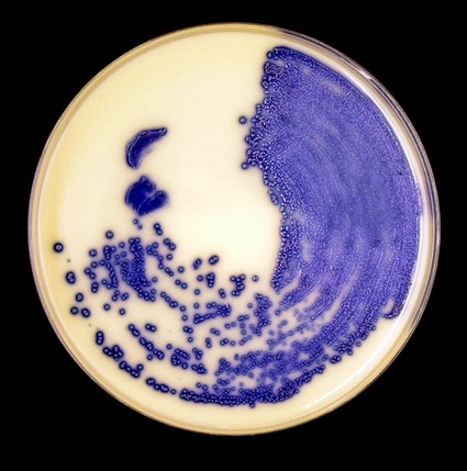
Enterococcus faecium on Oxoid ® Brilliance Chromogenic Media (24hrs at 37˚C)
Suspicious colonies are investigated further by determining the actual MIC of vancomycin using the E-test as mentioned above. Enterococci with MIC’s greater than 8 µg/ml are considered to be VRE. (Identifications can be confirmed using common microbial identification platforms or traditional tests).
Patients known to harbour VRE’s can be isolated and contact precautions implemented to reduce the likelihood of dissemination.
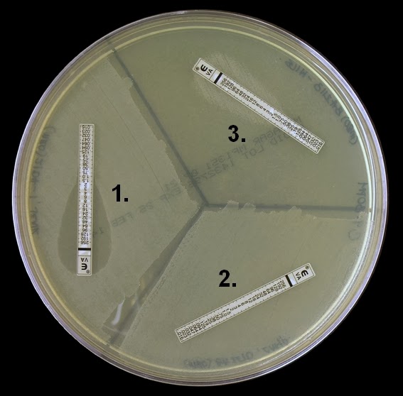
The antibiotic sensitivity plate above shows three different organisms subjected to an e-test in order to determine their susceptibility to vancomycin. Organism (1) is an enterococcus susceptible to vancomycin (VSE), (2) shows an enterococcus resistant to vancomycin (VRE), and (3) shows a curious response to vancomycin I had never before encountered. This organism exhibits vancomycin dependence! (VDE).
Note:the E-test is a strip impregnated with a continuously varying concentration of antibiotic along its length. On the Vancomycin E-test strip the concentration varies from 0.016 µg/ml to 256 µg/ml. The MIC value is where the growth/no-growth intersects the strip. The zone of inhibition is narrowest as it approaches the point of intersection and widest at the top of the strip where the concentration is the greatest.
Okay, what gives? Let’s dig deeper:
Vancomycin binds to the terminal D-Ala:D-Ala structure in the peptidoglycan layer of the enterococcal cell wall. This prevents the crosslinks from forming and the pentapeptide structures from extending during synthesis. Cell wall formation is terminated.
In both Van-A & Van-B genotypes, the gene cluster acts to a) detect the presence of vancomycin and start transcription of specific resistance genes, b) form and incorporate D-Ala:D-Lac into the growing peptidoglycan wall, and c) eliminate any D-Ala:D-Ala precursors, thereby eliminating the vancomycin sensitive pathway of peptidoglycan formation.
In other words, vancomycin binds to D-Ala:D-Ala, however by the enterococcus substituting D-Ala:D-Lac into the structure, vancomycin will no longer bind rendering the organism vancomycin resistant.
.jpg)
Vancomycin Dependence (VDE):
It has been proposed that vancomycin dependence may develop from the loss of a functional D-Ala:D-Ala ligase in the VRE strain, which is then unable to survive unless vancomycin induces the production of D-Ala: D-Lac ligase. This dependence involves mutations to the dll gene which encodes the enterococcal D-Ala:D-Ala ligase protein.
In other words, Vancomycin induction of the Van A or Van B ligase would compensate for the absence of the native ligase by producing D-Ala:D-Lac allowing for cell wall precursor synthesis. Since these ligases are only induced in the presence of vancomycin, the organisms cannot grow in the absence of this antibiotic unless it reverts to the vancomycin resistant form.
Revertant Mutant Enterococci:
If a particular strain of enterococcus becomes dependent on vancomycin for its growth and survival, it would seem logical that removing vancomycin would cause the organism to die. Surprisingly, this is not always the case as the organism may undergo a ‘revertant’ mutation. The enterococcus may undergo another genetic change that restores the D-Ala:D-Ala ligase function. The organism may enter a cyclical mutational change allowing it to shift between resistant and dependant phenotypes.
Withdrawal of vancomycin may not be adequate to eliminate vancomycin dependent strains.
On the first photograph of this post, the colonies randomly scattered throughout the agar surface, away from the E-test strip may be revertant colonies. These colonies were not apparent after 24 hours however these colonies appeared after sitting on the bench for approximately another 16 hours.
Revertant strains have not been observed in clinical situations and the presence of VDE does not appear to affect the patient’s clinical outcome.
These are the kind of microbiological oddities that give this blog its title “Fun With Microbiology”!