Quantcast
Channel: Fun With Microbiology (What's Buggin' You?)
Viewing all 85 articles
Browse latest View live

Iodamoeba bütschlii

$
0
0

Iodamoeba bütschlii(Intestinal Protozoa – Amoebae) Intestinal Parasite

Distribution:  Iodamoeba bütschlii can be found worldwide however it is less common than Entamoeba coli or Endolimax nana.

Pathogenicity:  I.bütschliiis considered non-pathogenic however it must be correctly identified to distinguish it from pathogenic intestinal amoebae.  Presence of these intestinal amoebae is evidence that the person carrying it has come in contact with a contaminated source, raising the possibility of the presence of other parasites.

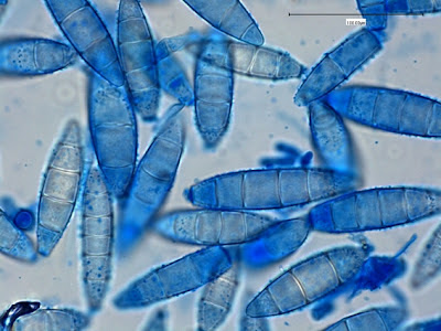
Trophozoites:Trophozoites reside in the large intestine where they survive by ingesting bacteria and yeast but not red blood cells.  In a direct wet preparation made from freshly obtained specimen, the trophozoites display a sluggishly progressive motility through hyaline pseudopodia.  In an iron hematoxylin or trichrome stained preparation, trophozoites exhibit a wide range in size, varying from 6 to 25 µm with the majority around 9 to 14 µm.  Cytoplasm has a coarsely granular appearance and food vacuoles containing ingested material may be visible.
The trophozoites have a single nucleus which is surrounded by a delicate nuclear membrane devoid of peripheral chromatin.  As such, a stained preparation may not delineate the nuclear membrane and the karyosome may simply appear to be contained in a vacuole.  The karyosome is large, irregularly rounded and may be central or somewhat eccentric.   With optimally stained preparations, granules surrounding the karyosome may be visible.  These chromatin granules may arrange to form a ring or radiate outwards between the karyosome and the nuclear membrane.
 
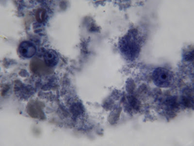
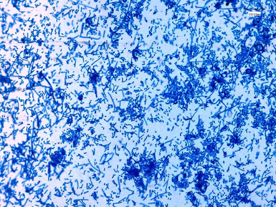
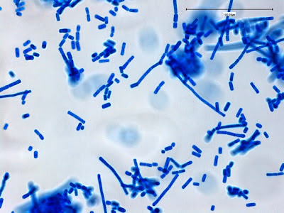
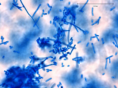
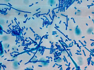
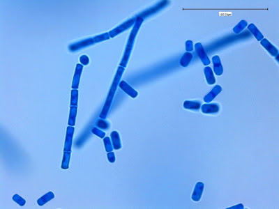
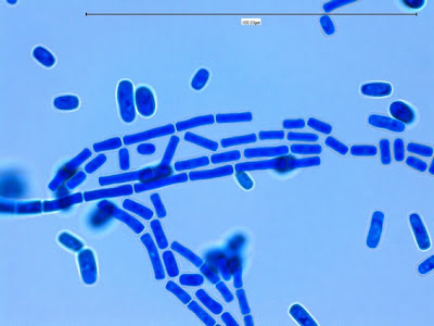
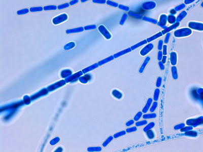
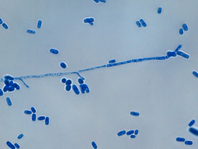
Note: Photos of  I.bütschlii trophozoites and cysts stained by using the Iron Hematoxylin stain follow.  I believe I took all at X1000 magnification.  Size may vary between photographs due to my cropping of the photographs.  Nikon Coolpix camera used for all but the last photograph.

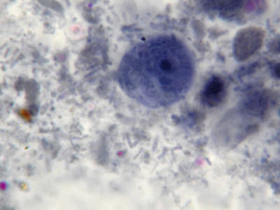
 I.bütschlii trophozoite (Iron Hematoxylin Stain X1000 Nikon)

Ditto

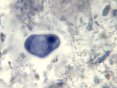
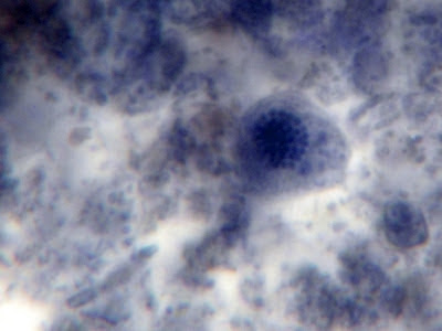
 I.bütschlii trophozoite showing large blotchy karyosome withing nucleus.  Nucleus outline visible showing no peripheral chromatin.

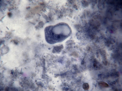
I.bütschlii trophozoite - cell shape is more irregular that Endolimax nana which is usually rounder.
 
 I.bütschlii trophozoite - again rather irregular shape.  Reddish spot (inclusion/surface?) is material which happened to stain acid-fast with the carbol fuschin staining stage added to detect the presence of Cryptosporidium parvum or Cyclospora cayetanesis oocyts if present.

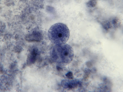
 I.bütschlii trophozoite - two cells.  Difficult to distinguish from E.nana trophs although generally more irregular in shape and space between nuclear membrane and karyosome appears clearer with fewer chromatin granules.

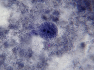
 I.bütschlii trophozoite - Binucleated cell

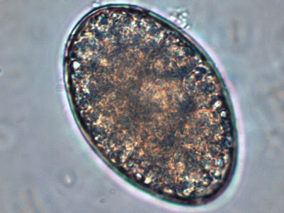
Cysts:  Cysts of I.bütschliirange in diameter from 6 to 16 µm, averaging 9 to 10 µm and may appear spherical however they are usually ovoidal or irregular in shape.  The most striking feature is the large glycogen vacuole which may contribute to half, if not more of the cysts size.  In fresh specimens stained with iodine, the glycogen vacuole appears yellow-brown to brown in colour.  It is because of this staining property with iodine thatIodamoeba acquired its generic name.  In an iron hematoxylin stained preparation, the glycogen vacuole appears clear or off-white’.  The cyst nucleus is large, often irregular in shape, with the karyosome usually eccentric in position.  It may even appear pressed against the nuclear membrane.  As with the trophozoites, the nuclear membrane is devoid of peripheral chromatin and therefore may not be visible.  In well stained preparations, chromatin granules may be form a crescent shape partially surrounding the karyosome. Linin fibrils may be seen running between the karyosome and the chromatin granules.  This arrangement when visible has been described as a ‘basket of flowers’, with a distorted karyosome forming the ‘basket’, the linin fibrils as the stems and the granules as the blossoms.  Unfortunately my staining and/or the resolution of the camera failed to pick up this fine detail in the photos which follow.

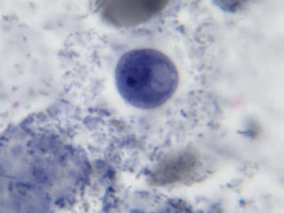
I.bütschlii cyst - Nuclear membrane all but invisible.  Large glycogen vacuole seen on right side of cell.

I.bütschlii cyst - large glycogen vacuole takes up most of the cell's interior.

Ditto

I.bütschlii cyst - irregular shaped cell with large glycogen vacuole.  Another view.

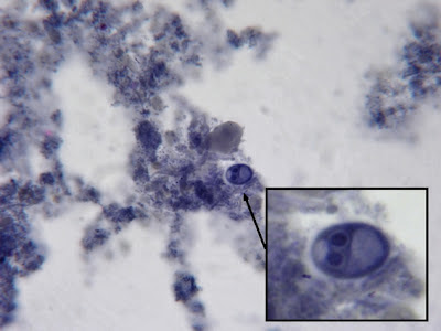
I.bütschlii cyst - binucleated cell.

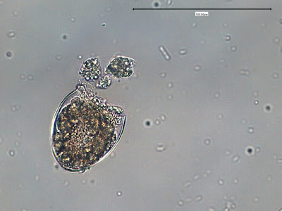
I.bütschlii trophozoite& cyst. Cyst nucleus not visible in this view.

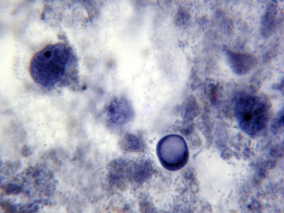
I.bütschlii trophozoite & cyst. Cyst nucleus not visible in this view.

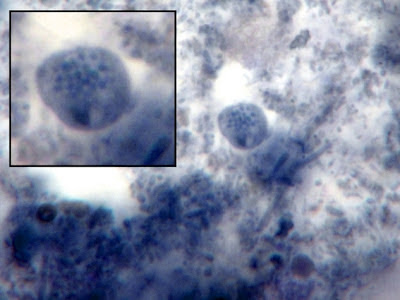
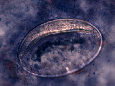
I.bütschlii - two cysts. 

 I.bütschlii -trophs & cyst

 I.bütschlii - Cysts in concentrate (unstained)
Trophozoites may be very difficult to recognize in an unstained concentrate as the nucleus is all but invisible.  The cyst is more obvious because of its large vacuole which, as above, appears as a void within the cell.  Iodine  can be added to stain the glycogen, making it more visible with the production of a dark brown colour.

Two I.bütschlii trophozoites seen and one Chilomastix mesnilii trophozoite (just below and to the left of lower I.bütschlii trophozoite)  This photo taken with the DMD-108 Microscope/camera.

Diagnosis:  Unstained preparations are inadequate for detecting I.bütschlii as the features are poorly defined with the nucleus often undetectable.  The cyst’s vacuole may be the only structure visible suggesting the presence of I.bütschlii.  Definitive diagnosis can only be made with the iron hematoxylin or trichrome stained preparation.  The cyst form is unique and cannot be mistaken for any other organism.  The trophozoites however may be mistaken for Endolimax nana trophozoites as the size range overlaps and both have a similar nuclear arrangement – a large karyosome (endosome) with no peripheral chromatin on the nuclear membrane.  E.nana trophs are generally smaller, having a less granular cytoplasm and the space between the karyosome and nuclear membrane may be devoid of the chromatin granules seen in I.bütschlii.   Differentiation may still be challenging.
A diarrheal sample is more likely to yield trophozoites rather than cysts.

Epidemiology, Prevention & Treatment:  Infective cysts are transmitted through the ingestion of contaminated food or water.  Infection is more likely where sanitary conditions are lacking.  As I.bütschlii is considered to be non-pathogenic, treatment is not recommended.


Aspergillus sydowii

$
0
0

Aspergillus sydowii(Fungus)

Distribution:  Worldwide distribution however may occur with increased frequency between 26o– 35o latitudes.  Primarily found in soils but has been recovered from a variety of sources, both indoor and outdoor.

Pathogenicity:Implicated as an agent of invasive aspergillosis, keratomycosis (infection of cornea/eye), and onychomycosis (nail infection).

  Aspergillus sydowii SAB 10 Days

Macroscopic Morphology: Growth rate is moderate.  Colour is influenced by media. It has been described as a blue-green to dark green to greyish-turquoise.  Colonies may have straw-coloured to reddish-brown shades with exudate.  Reverse is usually maroon, reddish, to reddish-brown.  The isolate presented here was grown on SAB media and appears dark green with a white fringe.  Texture has been described as lanose (woolly), velutinous (dense, silky hairy) or floccose (tufts of hair/hairy).  All the preceding means is that A.sydowii produces rather long conidiophore stipes (~stems) up to 200 µm which can give the colony that woolly or hairy appearance.

Micsroscopic Morphology:  The long, smooth-walled stipes which bear the conidiophores are hyaline generally (translucent/transparent/colourless) or slightly brownish. The vesicles (7.0 µm - 17 µm wide) may appear sub-spherical, pyriform (pear or teardrop shaped) to somewhat clavate (club shaped).   Conidiogenous structures are biserate with metulae[1](2 µm -3.5 µm by 4 µm -6 µm) and phialides (2 µm – 3 µm by 5 µm – 7 µm) in size.  Diminutive conidial structures are produced by many isolates which may resemble penicillate (like Penicillium) heads.  Conidia are spherical, echinulate or spinose (rough, jagged texture), and are about 2.5 µm to 4.0 µm in diameter.  Hülle cells may be present.

Note: All photos below were taken with the Leica DMD-108 digital microscope.  The optical magnification is recorded, however, if a +10 appears after the optical magnification factor, it denotes an additional 10% digital magnification.


 Aspergillus sydowii -Slide Culture 48 hrs (X250 LPCB)


 A.sydowii - Adhesive Tape Preparation (X1000 LPCB)

Aspergillus sydowii (X1000 LPCB)
Vesicle with biserate conidiogenous structures

Ditto
Aspergillus sydowii - Adhesive Tape Preparation (X1000+10 LPCB)
Vesicle bearing conidiogenous metulae & phailides (biserate)

A.sydowii (X1000 LPCB)
(Note 100 µm bar at upper right)

As above -another view (X1000+10 LPCB)

A.sydowii -Yet another view - As above

A.sydowii (X1000+10 LPCB)
(Note echinulate or spinose [rough] surface texture of conidia)

Aspergillus sydowii (X1000 LPCB)

Aspergillus sydowii - Long Stipes (X1000 LPCB)

A.sydowii - Adhesive Tape Preparation (X1000 LPCB)
(Note reduced conidiophore structure resembling Penicillium species)

A.sydowii conidia (X1000+10 LPCB)
(Note echinulate or spinose [rough] surface texture of conidia)

Note:  They may be distinguished from Aspergillus versicolor by their turquoise colony hue if grown on CYA25 agar.  The moderately growing A.sydowiiproduces rather small colonies within a week compared to many other Aspergillus species.  The rough conidia borne from small, compact biserate conidial heads on hyaline to slightly brown stipes is also distinctive.


[1]See A.fumigatus post for illustrated description of structures medulla & phialides

Microsporum gypseum

$
0
0

Microsporum gypseum(Fungus/dermatophyte)

Ecology & Pathology:Micrsporum gypseum is a geophilic (soil loving) dermatophyte which occasionally causes skin infections (tinea corporis) and/or scalp infections (tinea capitis). Onychomycosis (nail infections) have also been reported.  It has also been isolated from horses, dogs, cats and rodents.  A cosmopolitan fungus (found worldwide).

Macroscopic Morphology:  As with other fungi, colour is influenced by the media it is grown on.  Colony generally described as yellowish-buff to a dark cream or tan colour in colour.  The colony may develop a sterile white ‘feathered’ hyphal border or a cottony white raised center.  The reverse may be yellow, orange-tan or brownish-red in colour with possible pink to purplish tinges.  Colonies are generally flat with a granular or powdery texture and exhibit a moderately rapid growth rate, maturing in about one week.

Microsporum gypseum SAB at 7 Days (Nikon)

Microscopic Morphology:  M.gypseum has septate hyphae along which sessile or stalked clavate (club shaped) microconidia (3 – 8 µm X 2 -3 µm) may be found.  The fusiform (spindle shaped) macroconidia (8 – 15 µm X 22 – 60 µm) are relatively thin walled, verrucose (with bumpy surface) and contain about 3 – 6 internal cells.  M. gypseum’s macroconidia has a rounded apical end while the base is truncated and may show an annular frill.  They are usually produced in great numbers.  This differentiates M.gypseum’s macroconidia from the M.canis macroconidia which has rather pointed ends and the rather rare & distorted macroconidia produced byM.audounii.

Note: Photos that follow were taken with the Leica DMD-108 digital microscope.

 M.gypseum -Slide culture at 48 hours (X100 LPCB)

 M.gypseum -Slide culture (X250 LPCB)
Hyphae throughout, some showing microconidia, larger macroconidia at left of photo

M.gypseum -microconidia along hyphae are more visible in this photo. Several macroconida throughout. (X250 LPCB)

M.gypseum - another view of both macro & micro conidia (X250 LPCB)

M.gypseum macroconidia (X 400 LPCB)
(Note 100 µm bar at top right of photo)

M.gypseum macroconidia and sessile microconidia attached to hyphae
(X400 LPCB)

M.gypseum clavate microconidia (sessile & stalked?) attached to hyphae
(X1000 LPCB)

M.gypseum - numerous fusiform (spindle) shaped macroconida
(X400 LPCB)

M.gypseum - numerous macroconidia showing up to six internal cells in each.  Rough 'verrucose' surface is evident.  Adhesive tape preparation. (X1000 LPCB)

M.gypseum macroconidia - on left still attached to conidophore,  Rough verrucose surface on lower middle macroconida. (X1000 LPCB)

M.gypseum - more of the same, but I just love them!
(X1000 LPCB)

M.gypsium - macroconidia and a few microconidia (X1000 LPCB)

M.gypseum macroconidium showing rounded apical end and truncated end where once attached to conidiophore.  Six internal cells or compartments visible.  Rather thin walled with somewhat rough texture evident.  (X1000+10 LPCB)

M. gypseum - nearly identical to the macroconidia above, this cell also has six internal cells or compartments with verrucose surface.  Nature's own canoe!  (X1000+10 LPCB)
*  *  *
  Physiological Tests:  M.gypseumis positive for the hair perforation test.  It requires no special growth factors and produces no change in pH when inoculated into BCP-milk solids-glucose media.  It is urea positive.

 M.gypseum - Intended as computer wallpaper (1024 X 768 when posted)


Sphaerita spp.

$
0
0

Sphaeritaspp.

“Big bugs have little bugs, on their backs to bit them’
Little bugs have lesser bugs, and so on ad infinitum”
Anonymous

Some protozoan parasites can themselves be parasitized.  A hyper-parasite! The genus Sphaerita is considered to be a lower fungus and some species are capable of invading the cytoplasm of some amoeboid parasites.  Another parasite of parasites is Nucleophaga species which invades the nucleus.  Sphaerita, (sometimes called Polyphagaspp.) appear as tightly packed clusters within the cytoplasm and measure approximately 0.5 µm to 1.0 µm.

The parasite show below is possibly an Entamoeba coli, however the nucleus is not visible as it is out of the plane of focus.  Sphaerita appears as the small dots within the cytoplasm.

 An amoeboid parasite itself parasitized by the fungal parasite Sphaerita spp.  The Sphaerita appear as the neatly arranged circular dots withing the cytoplasm.
 Iron hematoxylin stain (X1000) Nikon


 An amoeboid parasite itself parasitized by the fungal parasite Sphaerita spp.  Doesn't appear to be digested material or inclusions. This parasite appears to have a rather dark blotchy karyosome reminiscent of Iodamoeba  bütchlii.  Unfortunately I failed to record measurements when I took these photos.  E.coli, Iodamoeba  bütchlii and E.nana were all present in this particular specimen.
Iron hematoxylin stain (X1000) Nikon

  Return Home (most recent posts)

Geotrichum candidum

$
0
0

Geotrichum candidum  (yeast/fungus)

Ecology:Ubiquitous worldwide distribution.  Has been isolated from soil, water, sewage, cereals, dairy products and various plants.   Geotrichum species have been considered a part of normal commensal flora when isolated from the sputa and/or feces of health humans.  The genus Geotrichum has several species with Geotrichum candidumbeing the most common.

Macroscopic Morphology:   On SAB, colonies exhibit moderately rapid growth, producing off-white to cream coloured colonies with a butyrous texture with a velvety, suede-like or ground glass/matt appearance.  Colonies grow best at around 25oC to 30oC however growth may be restricted at 37oC.
 Geotrichum candidum on SAB agar incubated at 30oC for 5 days

Microscopic Morphology:  Geotrichum species produce hyaline (clear), septate hyphae which show dichotomous branching (7µm – 11 µm wide).  Advancing undifferentiated aerial hyphae produce chains of arthroconida which fragment into individual cells of variable size (6 -12 µm to 3 – 6 µm).  Cells can be cylindrical in shape or may become barrel shaped.  Blastoconidia, conidiophores and pseudohyphae are not produced by Geotrichum species.  Disjunctor cells (empty cells in between arthroconida that fragment to release the arthroconidia) are absent which differentiates them from Coccidioides immitis and Malbranchea.  . Blastoconidia, conidiophores and pseudohyphae are also not produced.

Note: Photos taken with the Leica DMD-108 Microscope.  (a +10 after any magnification indicates another 10% digital magnification factor in addition to the optical magnification of the objective.

 Geotrichum candidum  at ~24 hrs (X250) LPCB

 Geotrichum candidum  at 48 hrs (X250) LPCB

 Geotrichum candidum   (X400) LPCB

Ditto

Geotrichum candidum showing hyphae, arthroconidia in chains and individual arthroconidia from the fragmentation of the chains  (X400) LPCB

Geotrichum candidum showing chains of arthroconida and separate arthroconidia
(X1000) LPCB

Geotrichum candidum showing chains of arthroconida and separate arthroconidia
(X1000+10) LPCB

 Geotrichum candidum showing chains of arthroconida, individual arthroconida from the fragmentation of the chains and septate hyaline branching hyphae in the lower right of photo.
(X1000+10) LPCB

Geotrichum candidum - yet another view to get a feel of the organism,
(X400) LPCB

Geotrichum candidum - showing hyaline hyphae with some rudimentary branching and evidence of the development of arthroconidia as chains towards the right.  Individual arthroconida appear throughout photo.  (X400) LPCB

Geotrichum candidum - individual arthroconidia.  Some of the arthroconidia appear as cylindrical with rounded ends.  Some arthroconidia look somewhat barrel-shaped with some variation in size.  Unsure of the round cell near the middle (X1000+10) LPCB

 Pathogenicity:  Geotrichumspecies is the causative agent of geotrichosis.  Broncheal and pulmonary infections are the most frequently reported form of the disease, particularly in the immunocompromised host.  Oral, vaginal, cutaneous and alimentary infections have also been reported.

Geotrichum species (candidum) showing dichotomous branching and chains of arthroconida which are fragmenting into individual arthroconidia.  Some cells appear with rather square ends and some rather round.  (X1000+10) LPCB
This photo intended as computer wallpaper (1024 X 768) when posted.


Diphyllobothrium latum (revisited)

$
0
0

Diphyllobothrium latum(Helmith/Cestode)

Note:  This post is entitled Diphyllobothrium latum "Revisited" as I posted a photo several years ago when a fixed film camera was the only tool in my arsenal.  I'm sure you'll find these new photos superior.  To visit the old post click below;

Known as ‘Fish Tapeworm’ or ‘Broad Tapeworm.  

Disease:  Causes’ Diphylobothriasis’.  In some instances the infection can be relatively harmless while in others it results in an illness resembling pernicious anemia.  The tapeworm may be depriving the infected host of vitamin B12 as the adult worm contains a high concentration of this vitamin.  Disease is acquired by eating raw or insufficiently cooked fish.  Campers who cook their catch of fish over an open fire in the wild and cultures who eat raw or pickled (incomplete) fish may be more likely to acquire the infection.  Lives in the proximal portion of the jejunum.

Geographic Distribution:  Found in temperate regions with cold clear lakes.  Prevalent in Japan, Russia, Scandinavia. Finland, the Baltics as well as Canada and Alaska.   Has been found in the Great Lake Regions of the United States however is not considered to be endemic.   In other regions of the world, different species of Diphyllobothrium may infect humans and susceptible mammals.

Life Cycle:  Diphyllobothrium latum required two intermediate hosts in order to complete its life cycle.  Briefly, the eggs are passed from feces which then hatch into small ciliated coracidium larvae.  These coracidia survive until they are ingested by copepods where the second larval stage (procercoid) develops with growth.  These fresh water crustaceans are themselves ingested by fish where they continue to grow where they develop into the plerocercoid stage.  Small infected fish may not be suitable for human consumption however they in turn may be ingested by larger fish thereby infecting them.  The larvae invades and resides in the flesh of the fish.  As the larger fish consumes smaller infected fish, the infectious potential increases when consumed by humans.  The plercoceroid larva (or sparganum) is not digested but remains in the small intestine where it develops to an adult tapeworm in about three to five weeks. The tapeworm, unless treated, may live for 25 years or longer.

Diagnosis:Infection with Diphyllobothrium latum is made by demonstrating the ova (eggs) in feces.  Occasionally segments of the proglottids (broken off segments of the worm) can also be found in the feces.

Ova (Eggs):  The Diphyllobothrium latum ova are ovoid in shape and have an operculum.  (a small cover or hatch through which the larvae can escape).  The yellowish-brown eggs are moderately thick-walled and are usually about 58 – 75 µm by 40 – 50 µm in size.  They may have a small know at the end opposite of the operculum however it may be indistinct.  The egg is unebryonated when passed in the feces.

Adult Worm:  The clinical laboratory usually encounters the egg rather than the adult worm as it is the stool (feces) which is usually submitted for diagnosis.   As I have no adult worm specimen, I’m unable to present any photos.  Consult other sources for photos of the worm which may grow to 4 – 10 meters in length.

 Two Diphyllobothrium latum eggs (arrows) in fecal concentrate (X100 Nikon)

 Two Diphyllobothrium latum eggs in fecal concentrate (X250 Nikon)

 Ditto

 Diphyllobothrium latum egg.  Arrows point to the the operculum or 'hatch' through which the larvae can escape. The protuberance, which can be somewhat inconspicuous, usually appears as a slight 'point' or 'bump' on the end opposite of the operculum. It is not evident on any of the photos in this post.  It does appear in my 2008 D.latum post. Fecal concentrate. Click on photo to enlarge for better viewing.
(X250 Nikon - size differs from previous photos due to cropping)

Broken Diphyllobothrium latum egg.  This is not an open operculum but rather a broken egg.
(X400 Nikon -Fecal Concentrate) Note micron bar in upper right.

Diphyllobothrium latum egg (X1000+10* -DMD-108: Fecal Concentrate)

Diphyllobothrium latum egg (X1000+10 DMD-108 Hematoxylin Stain)

Diphyllobothrium latum egg (X1000+10 DMD-108 Hematoxylin Stain)
(appearance differs due to perspective/orientation of egg)

Diphyllobothrium latum egg (X1000 DMD-108 Hematoxylin Stain)
(appearance differs due to perspective/orientation of egg)
Note micron bar in upper portion of photo

Diphyllobothrium latum Computer Screen Wallpaper (1024 X 768 when posted)
Ova in fecal concentrate X1000+10*DMD-108

*DMD-108 X1000 optical magnification (objective) + X10 digital magnification.


*   *   *

Paecilomyces lilacinus

$
0
0

Paecilomyces lilacinus(Fungus)

Ecology:  Wide-spread cosmopolitan saprobe of soil and decaying plant material.  Paecilomycesspecies have been implicated in the spoilage of food.  Has been isolated from cosmetics and some species of Paecilomyces parasitize insects.  Grows well at 30oC but growth is restricted at 37oC

Macroscopic Morphology:  As with many fungi, colour is influenced by the media it grows on.  Colonies often display a faint violet or mauve colouration (hence the name derived from lilac) which may develop to a reddish-grey tint.  The reverse is unremarkable.  Colonies are woolly to floccose and growth is moderately rapid, reaching 3 – 4 cm in about a week.


 Paecilomyces lilacinus on SAB ~2 weeks at 30oC - Note slight lilac (purplish) colour.

Microscopic Morphology:  P.lilacinus produces hyaline (clear), septate hyphae.  Conidiophores stipes (3 -4 µm wide) usually arise from submerged hyphae and can achieve lengths between 400 µm – 600 µm.  Branched conidiophores give rise to clusters of phialides.  The phialides have a broader base which tapers to form a long, narrow neck giving a delicate elegant appearance if compared to Penicillium.  The phialides tend to curve away from the central axis of the conidiophore.  Conidia (2.5 – 3.0 µm by 2.0 – 2.2 µm) form at the apical end of the phialides and are ellipsoidal (spindle) to fusiform in shape.  They arise in rather long divergent chains which are easily disrupted.

 P.lilacinus LPCB (X400 -Nikon)

Ditto


 More of the same in order to give a sense of how P.lilacinus appears.  Delicate brush-like appearance with phailides turning away (outward) from stipe). Kind of resembles Neptune's trident! Conidia in chains are copiously produced and easily disrupted.  I found no advantage with either adhesive tape preparations or slide cultures in preserving the appearance.  (LPCB X 400 Nikon)

 P.lilacinus - another view of conidiophores and chains of conidia (LPCB X1000 I believe, DMD-108)
P.lilacinus - I hate books that offer one B&W photo or a simple line drawing as the sole example of a particular fungus.  I find it difficult to get a real sense of the organism.  So I tend to overdo it in these posts by offering a number of views.  (LPCB X1000 Nikon)

P.lilacinus - showing a long chain of conidia which are delicate & easily disrupted
(X1000+10 DMD-108)

P.lilacinus - showd delicate, elongated, flask-shaped phailides bending away (splaying out) from stipe. (LPCB X100, DMD-108)

P.lilacinus - final view of phailides and chaining conidia.
 Size difference from previous photos is due to cropping.
(LPCB X1000 Nikon)

Pathology:  Often considered an environmental contaminant however should not be immediately dismissed as such.  P.lilicinus has been isolated from ocular infections such as keratitis, endopthalmitis and corneal ulcers.  Also has been implicated in bursitis, sinusitis, endocarditis, pulmonary infections, cutaneous infections, catheter related infections and even onychomycosis (nail infections).  Diabetic patients and immunocompromised patients may be at greater risk of infection.  P.lilacinus been successfully treated with ketoconazole.


*   *   *

Fusarium oxysporum

$
0
0

Fusarium oxysporum (Fungus)

Ecology:
Fusarium oxysporum has widespread distribution and can be isolated from soil and plants and decaying vegetative material (saprobe).  It is a phytopathogen (plant pathogen) of many species.

Macroscopic Morphology:
On Sabouraud-Dextrose (SAB) media (30oC) this isolate of F.oxysporum grew rather rapidly to produce of-white floccose (cottony) colonies with the aerial mycelia becoming tinged in purple.  The reverse was a rather non-descript pale to yellow.  Other sources have described the reverse as having a purple colour, particularly on Potato-Dextrose agar, a characteristic not seen on the isolate presented here and grown on SAB.


Microscopic Morphology:
Hyphae are hyaline (clear/non-pigmented) and are septate (show divisions or walls within the hyphae).  Conidiophores are rather short (8 - 14 µm) and usually non-septate when compared to other Fusarium species. The conidiophores have a somewhat inflated appearance as their sides aren’t parallel but slightly bulge out slightly in the middle.  These conidiophores (phialides, or more accurately monophialides) are produced singly as they extend from the aerial mycelium.  Microconidia (5 - 12 X 2.3 – 3.5 µm) are usually non-septate, ellipsoidal and are straight or slightly curved in shape as they are abundantly produced from the tip of these phialides.  Microconidia are produced singly and never in chains.  These microconidia may accumulate around the tip of the phialide if not dispersed (false head).
Macroconidia (23-54 X 3.0 – 4.5 µm) are fusiform in shape (hence the genus name), and have a slightly pointed apical tip with a basal ‘foot’ cell (pedicellate) at the opposite end.  The fusiform macroconidia are also somewhat curved making it appear sickle-shaped or perhaps canoe-shaped in profile.  They usually contain 3-5 compartments or divisions within the macroconidium.
Smooth-walled terminal or intercalary chlamydospores (5 – 13 µm dia.) may be found.

 Fusarium oxysporum - First look at low power (X100 LPCB)

Fusarium oxysporum (X400 LPCB: DMD-108)

Fusarium oxysporum (X400 LPCB: Nikon)
Microconidia can be seen accumulating around the tips of the phialides (see below).

Fusarium oxysporum (same photo as above but cropped)
Arrows point to tips of phialides where microconidia are produced and accumulate

Fusarium oxysporum - Microconidia and a few Macroconidia (X100 LPCB: Nikon)

Fusarium oxysporum - again showing hyphae from which monophialides extend, producing microconidia at the tips which can accumulate there unless dispersed.
 (X1000 LPCB (DMD-108)

Fusarium oxysporum - another view (as above) - Note micro bar at top of photo.
 ( X1000 LPCB: DMD-108)

Fusarium oxysporum - yet another view of two parallel hyphae from which the phialides extend producing micro (&/or macro) conidia.  Conidia vary in shape from the rather straight fusiform (lens shaped) to the curved banana or canoe shape.
 (X100 LPCB: DMD-108)

Fusarium oxysporum - microconidia accumulating at the tips of phialdes
(X1000 LPCB: DMD-108)

Fusarium oxysporum - microconidia in various stages of development (arrows) at the tips of the monophialides from which they are produced.
 (X1000+10* LPCB: DMD-108)

Fusarium oxysporum - (yeah, I like photos) -again, microconidia accumulating around tips of phialides (X1000+10* LPCB: DMD-108)

Fusarium oxysporum - somewhat inflated (sides not parallel) phialides extending from hyphae where conida are produced (arrows).  The one on left has separated from the tip of the phialide whild the one on the right is young and still developing.
  (X1000+10* LPCB: DMD-108)

Fusarium oxysporum - micro & macro conidia accumulated along side of hyphae.
(X1000+10* LPCB: DMD-108)

Fusarium oxysporum - slightly curved micro & macro conidia pictured
(X1000+10* LPCB: DMD-108)

Fusarium oxysporum - chlamydospore present (arrow).  Difficult to see from the orientation but I believe this was an intercalary chlamydospore (growing between the hyphae and not a terminal chlamydospore at the end of a hyphae.
  (X1000+10* LPCB: DMD-108)

Fusarium species - Just for comparison of the shape.  The photo on the left is of micro (and a few macro) conidia produced by Fusarium oxysporum while the one on the right is of microconidia produced by Fusarium dimerum.  The F.dimerum is showing a greater curved "banana" or "canoe" shaped microconidia thatn the F.oxysporum. (Magnification not noted: Nikon for both)

Pathogenicity:
Infections with Fusarium species (fusariosis) may be localized or become disseminated.  Fusarium species are frequent agents of mycotic eye infections, particularly the cornea (keratomycosis, endopthalmitis).  They have also been implicated in onychomycosis (nail infections), catheter infections, peritonitis, sinusitis and septic arthritis.  As with many other fungi immunocompromised and neutropenic patients may be at greater risk.  Fusariummay contaminate stored grain where some species can produce potent mycotoxins.  Food prepared from these contaminated grains may cause illness on ingestion.  Fusarium species may also be found as laboratory contaminants but must not be dismissed outright without further investigation.

Differentiation:
Fusarium oxysporumcan be differentiated from F.solanicomplex which produce thick, blunt macroconidia and long, narrow mono-phialides as well as numerous rough-walled chlamydospores.  The colonies, however, may appear brownish, particularly on Potato-Dextrose agar   F.oxysporum can be differentiated from F.verticillioides as this species has candle-shaped mono-phialides as well as baton-shaped microconidia in chains that are not easily disrupted.  Colonies however may appear purplish as those of F.oxysporumF.proliferatumalso is similar in appearance however produces polyphialides (many phialides as opposed to the single phialide in monophialides) from which delicate chains of microconidia can extend.  Macroconidia are seldom seen in most F.verticilliodesor F.proliferatum species unless induced by special media or exposed to UV light.

*DMD-108 microscope/camera is capable of taking photos with 1000X optical magnification plus and additional 10% digital magnification.


Adhesive Tape Preparation for Examination of Fungi

$
0
0


An earlier post showed how to make a Slide Culture of a Fungus for examination.  In this post I'll show how to make an adhesive tape preparation of a Fungus for examination.

Adhesive Tape Preparation or Slide Culture - which should I make?  Well, try both if you can.  I find that sometimes one may be superior to the other when trying to capture the structure of a particular fungus.

 The adhesive (sticky) tape preparation pulls up the structures and sort of locks them in place -stuck to the tape.  Or, it may be a bit more disruptive and perhaps destroy or scatter some of the fungal features.  You never no for sure until you try.  The other issue is the clarity of the tape itself.  Adhesive tape has the glue laid down on one side to make it sticky.  The evenness of the glue as well as the transparency of the tape can affect the quality of the image when viewed.  Adhesive tape is manufactured specifically for use with mycological specimens however I've found that any good commercial adhesive tape, such as Scotch Tape™, is quite adequate.  It should be obvious but I'll say it anyways: use only clear tape -do not use 'frosted' tape!

The advantage of a slide culture is that the fungus can attach itself to the coverslip as it grows and if removed very carefully, the features will not be disturbed and can be viewed as they naturally occur.  The trick is to be very gentle when removing and mounting the cover slip.

Well, nothing could be simpler:  Just grow your fungus on appropriate media and take a sample.  Structures may develop over time so you may wish to make adhesive tape preparations of the same fungal colony over several days.  Structures may also deteriorate or disappear on prolonged incubation so timing is important in making both adhesive tape preparations and slide cultures.

Important!!! Make all preparations within a biological safety cabinet (BSC) rated at a Level 2.

Here we go:
1. Get your supplies together.  You will need some adhesive tape (1).  Pictured here is a roll of  'Fungi-Tape manufactured by a laboratory supply company specifically for this purpose.  Also shown is a roll of Scotch™ brand transparent tape which is just as good.  You want to select a tape that is about the width of the slide.  Of course you will need a microscope slide (2) and some Lactophenol Cotton Blue (LPCB).  This can be made up in the lab (formula at the end of this post), or can be bought "ready to use" from laboratory supply companies.  Pictured here is an ampule of LPCB (3) manufactured by Becton-Dickenson. The squeeze dropper is a quick and clean way of dispensing LPCB to the slide.

2. Dispense a line of LPCB on the slide.  The amount will dispensed will come with practice.  Too much and the tape will float with the LPCB oozing out the sides.  Too little and any spores present will not come in contact with the LPCB, posing a possible contamination problem.  I like to stick one end of the tape to my thumb and the other end to my middle finger.  (admittedly rather difficult to see in this photo due to the cramped confines of the BSC.)

3. This allows me to use my free index finger (pointer finger) in between my thumb and middle finger to push the sticky side of the tape down onto the colony.

4. Uncover petrie dish containing fungus and  prepare to take a sample.


5. With the tape ends stuck to your thumb and middle finger, press down with your index (pointer) finger to sample the fungal growth.  If possible, I try to get a sample from the middle of colony out to the edge, thereby sampling various ages of the colony. (younger growth on the outer edge).

6. As you remove the tape from the fungal colony, a representative sample of the growth should remain stuck to your tape.

 7. Line up and place the tape onto the slide where the LPCB should spread out to the edges of the tape.  This is somewhat tricky.  If you don`t manage to place the tape down exactly parallel to the edges of the slide, the tape will overhang the glass slide.  Spores loosely adhering to the tape may present a contamination hazard and the tape may stick to the microscope stage compromising its movement.  Removing and reapplying can be done but may further disturb the structures on  the tape.


8.  The adhesive tape preparation slide is ready to be viewed on a microscope (boy, I didn`t make a nice and even looking preparation for this photo, did I! - but you get the idea.)  I`m both making the preparation and taking the photos so both hands are full.

With a bit of skill, you can also use sticky tape to sample a fungus growing in a test tube.

1. Wrap tape into a circle, sticking one end to the other and then sticking the loop (arrow) to a wooden applicator stick or other long thin item.  Carefully insert the adhesive tape loop into the test tube culture media and press against the fungal growth.  Pull the applicator stick with the tape out of the test tube.  Going in and out takes patience and practice for if you touch the glass neck of the test tube with the tape, it will no doubt stick to the glass and your attempt will be ruined.

2. Once outside, carefully cut the tape loop open with scissors and place tape onto the microscope slide with LPCB.

Disinfect your scissors and dispose of all used materials in a safe manner!!

Lactophenol Cotton Blue

What is and why use Lactophenol Cotton Blue?

Well, if you wish to look as a fungus microscopically you no doubt want to enhance the features while doing it safely.  Lactophenol Cotton Blue (LPCB) is a mounting fluid used by most laboratories examining fungi.  It has several components/properties that make it ideal for this purpose;

  • Lactic Acid acts as a clearing agent and helps preserve the fungal structures
  •   Phenol kills the fungus making it safe to remove your slide preparation from you biological safety cabinet (BSC).  Spores are often abundant and can easily infect the mycologist or contaminate the laboratory if not killed.
  •   Glycerol is slightly viscous and prevents drying of the prepared slide specimen.
  •   Cotton Blue is an aniline dye which adds colour to the fungal preparation thereby enhancing and contrasting the structures.
 LPCB is sold commercially[i]in various volumes.  Pictured in the adhesive tape preparation photographs is a single use dispenser of LPCB.  A glass ampule is encased in a crushable plastic dispenser.  Crushing the glass held within the plastic sleeve by squeezing releases the LPCB to be applied where required.

LPCB consists of;


1.       Gently heat the phenol crystals (if used in place of the concentrated sol’n) in the lactic acid,   glycerol and water.
2.       Add cotton blue to the solution above
3.       Mix well and cool.
Notes:
Cotton Blue = Poirrier’s Blue
Solution can be stored at room temperature and dispensed with a pipette when needed.
10% Potassium Hydroxide (KOH) can also be used in some situations as a clearing agent which is lethal to fungi.  Obviously structures will appear clear.


[i]The LPCB pictured in these posts is manufactured by Becton-Dickinson.

Gliocladium species

$
0
0

Gliocladium species(Fungus)
Ecology & Pathogenicity:
Gliocladium is a cosmopolitan fungus which can be isolated from soils as well as decaying plant material (saprobe).  Gliocladium is considered to be a contaminant when found in the clinical situation as no cases of human or animal infections have been documented.

Colony Morphology:
Gliocladiumexhibits rapid growth, maturing within 4 to 5 days.  Surface growth is white to cream at first but quickly develops various shades of green.  The outer fringe may remain white. Some strains may also exhibit pink colouration on the surface.  Surface texture has been described as cottony to woolly.  Reverse is unremarkable, pale or slightly yellow in colour.

 Gliocladium species on SAB at 72hrs, 30oC

Microscopic Morphology:
Gliocladium produces hyaline (clear/non-pigmented), septate hyphae.  Extending from the hyphae are conidiophores which branch in the upper portion somewhat resembling Penicillium species.  Extending from the branching conidiophores are somewhat ‘flask-shaped’ phialides which are arranged in brush-like clusters at the apices.  The unicellular conidia produced by the phialides are smooth walled and ovoid in shape.  Conidia do not chain but rather accumulate at the apex of each individual conidiophore as a rather large slimy ball.

*   *   *
Gliocladium sp. on Slide Culture (X100 LPCB: DMD-108)

Gliocladium sp. -'balls' of conidia visible at top of phialides
(X400 LPCB: DMD-108)

Gliocladium sp. (X400+10* LPCB: DMD-108)

Gliocladium sp. - yet another view of hyphae, conidiophores, phialides & balls of conidia
(X400+10* LPCB: DMD-108)

Gliocladium sp. - and another view as above.
 (X400+10* LPCB: DMD-108)

 Gliocladium sp. - a few phialides without conida shows there somewhat "flask-like" or "inflated" shape. (X400 LPCB: DMD-108)

Gliocladium sp.  a good view of a conidiophore from which extend the brush-like phialides which produce the conidia.  (X400+10* LPCB: DMD-108)

Gliocladium sp.  This photo better shows what have been described as "flask-shaped" phialides
(X400+10* LPCB: DMD-108)

Gliocladium sp. -As above but a closer view.
(X1000+10* LPCB: DMD-108)

Gliocladium sp.  -More is better!  Another view to get a feel of what the phialides look like.
(X1000+10* LPCB: DMD-108)

 Gliocladium sp.  -a good shot of the brush-like phialides extending from the conidiophore and the "sticky" ball of conidia which have formed at the tips of the phialides but because of their stickiness, have remained together at the apex.  (X1000+10* LPCB: DMD-108)

 Gliocladium sp.  -Taking a step back, this photo shows the branching conidiophore, each bearing the brush-like 'Penicillate' phialides and ball of conida. (X400 LPCB: DMD-108)

Gliocladium species (X1000+10* LPCB: DMD-108)

 Gliocladium species (X1000+10* LPCB: DMD-108)

 Gliocladium species -large collection of conidia at apex of phialides
(X1000+10* LPCB: DMD-108)

 Gliocladium species - Intended as computer wallpaper (1024X768 when posted)

* The DMD (Digital Micro imaging Device) is capable of adding an additional 10% digital magnification to the optical magnification of the objectives


Exophiala dermatitidis

$
0
0

Exophiala dermatitidis (Wangiella dermatitidis)Fungus

Note: The current name for this fungus is Exophiala dermatitidis, changed fairly recently from Wangiella dermatitidis & Phialophora dermatiditis earlier still). Molecular studies have shown it is related to Exophilia as a separate species and does not warrant its own genus (Wangiella) containing the single species (dermatitidis). Originally it was thought this organism differed from Exophialaby its ability to produce phialides from which the conidia are produced.

Ecology: Exophiala dermatitidis is a cosmopolitan (found almost everywhere) saprobe (lives of off dying/decomposing material). It can be isolated from soil, decaying wood, plants and from water.

Macroscopic Morphology: Shown here on Sabouraud Dextrose Agar (SAB or SDA) incubated at 30oC, colonies are black to olive-black, often with greyish to brownish colours showing through, particularly at the periphery where the growth is younger. The brownish pigment may diffuse into the surrounding media.  It somewhat resembles molasses or perhaps dirty old motor oil. Reverse is much the same with a dark grey to iron-black colour. Growth is slow to moderate as the organism visible growth appears quickly but the colony is rather slow to expand and mature.  A distinguishing feature is the sticky, wet, mucoid growth this fungus initially produces.  A slimy string can be produced between the colony and an inoculating loop attempting to pick it up.  Some filamentous growth may occur on prolonged incubation or after several subcultures.  One source suggests that SAB media may encourage this.

Hints:  I didn’t have much luck with adhesive tape preparations with this ‘goo-y’ growth. It’s like trying to pick up a drop of motor oil with scotch tape!  Initially the fungus produced only yeast cells however some filamentous growth was finally visible at the center of the colony (oldest growth) after prolonged incubation (1 month).  Other sources suggest the velvety or filamentous growth may be first seen at the periphery.  The plate photo below is of a subculture incubated for about one month at 30oC.  The majority of the photos shown below were taken of Exophiala dermatitidisgrowing on a slide culture, harvested after about 10 days of incubation at 30oC.  I found this to be the most effective technique for viewing this organism. Hyphae are sparse but can be found growing out of the mass of yeast cells.

 Exophiala dermatitidis on SAB after about 1 Month at 30oC
This organism grows fairly slowly and produces a wet, sticky, rather black looking colony.  Reverse (not pictured) is much the same.  Hyphal growth may appear after prolonged incubation and perhaps  is encouraged by repeated subculture.  Note the center of the growth appears more dry. The colony above was so shiny and glistening from the wet appearance that white bright spots on the left side of the colony are simply reflected light. (Nikon)

Microscopic Morphology:  Yeast cells are abundant with early growth appearing hyaline (clear) but acquire an olive colouration quickly. The sparse hyphae are septate and also have a pale olivaceous appearance in a wet mount. Conidiophores are poorly differentiated, often indistinguishable from the vegetative hyphae.  Phialides are also poorly differentiated and are generally ellipsoidal in shape.  Exophiala dermatitidis may somewhat resemble Phialophorahowever they do not produce the collarettes that are present on Phialophora.  The phialides can be intercalary (produced along the hyphae) or terminal (at the end).  Conidia are smooth, one-celled, subglobose to ellipsoidal (2.5 - 4 µm by 2 – 3 µm).  Somewhat spherical phialides may also be present producing phialoconidia of about the same size.  The conidia generally accumulate around the apex (tip) of the phialide.

 Exophiala dermatitidis - Not much detail here but this photo shows the yeast cells mass at the bottom of the photo from which hyphae have grown outwards. This was taken from the growth adhering to a microscope cover-slip taken from a slide culture after 10 days incubation at 30oC.
(LPCB DMD-108 X40)

 Exophiala dermatitidis - as above (LPCB DMD-108 X250)
Just to give you an initial view of the colony before cranking up the magnification.

 Exophiala dermatitidis - at higher magnification the typical structures now appear.  At the bottom of the photo you can see the conidiogenous cells with conidia that have accumulated around their tip.  The cells at the top of the photo are out of the plane of focus but show much the same.
(LPCB DMD-108 X400)

Exophiala dermatitidis - Branched hyphae visible from which the poorly differentiated conidiogenous structures extend.  Oblong phialides can be seen and numerous oval conidia can be seen, particularly gathered around the somewhat obscured phialides on the left of the photo.
(LPCB, DMD-108 X1000)

 Exophiala dermatitidis- Elongated ovoid conidia seen gathered at the tip of one hyphal element and along it's length.  Inset & arrows: show the attachment point along the hyphae.
(LPCB, DMD-108 X1000)

 Exophiala dermatitidis - Shows hyphae which have poorly differentiated conidiogenous cells at the tip (ie. they don't look much different from the hyphae itself.  The phailides are the terminal cells from which the conidia are produced.  Again the conida can be seen accumulating around the tip of the cell from which they were produced.  Production of conidia can be seen at the end (terminal phialides) and along the length (intercalarly phialides).
(LPCB, DMD-108 X1000+10*)

 Exophiala dermatitidis- Hyphae seen with oval or 'vase' shaped intercalary phialides at various locations along it's length.  Again the far end of the hyphae shows poorly differentiated conidiogenous cells with a terminal phialide.  Conidia accumulate as clusters around both.
(LPCB, DMD-108 X1000)

 Exophiala dermatitidis- Another view shows hyphae with conidiogenous cells branching off and the somewhat oval of 'vase' shaped phialides at their apex (tip).  Elongated, oval conidia have accumulated around the tip.
(LPCB, DMD-108 X1000)

 Exophiala dermatitidis - Yeast cell mass at right of photo with single hyphae in center showing accumulation of oval conidia round the tip and along it's length.
(LPCB, DMD-108 X400+10*)

 Exophiala dermatitidis - Septa in hyphae are visible.  Phialide extending from hyphae is seen with conidia accumulated around the apex.
(LPCB, DMD-108 X1000+10*)

 Exophiala dermatitidis - Another view of a branch from a septate hyphae which can be seen differentiating towards the end where the conidia are accumulating  (ie. the cells are taper inwards where their are septa - at least two are visible)
(LPCB, DMD-108 X1000)

Exophiala dermatitidis - Mass of yeast cells seen at right of photo with two phialides seen extending from hyphae near center and the conidia they have produced.
(LPCB, DMD-108 X1000)

 Exophiala dermatitidis - Hyphae with phialides showing accumulated oval conidia around their tip.
(LPCB, DMD-108 X1000+10*)

 Exophiala dermatitidis - Another example as in the previous photo.  Compare this to Phialophora verrucosa.  No collerette is produced byExophiala dermatitidis but can be found on Phialophora.
(LPCB, DMD-108 X1000+10*)

  Exophiala dermatitidis - Yeast cells on left with hyphae extending outwards.  One phialides along length and an other(s) at end where conidia have accumulated at tip.
(LPCB, DMD-108 X1000+10*)

 Exophiala dermatitidis - Another photo, this with micron bar for scale.  Both Terminal (T) and intercalary (I) phialides are seen with the characteristic accumulation of conidia.
(LPCB, DMD-108 X1000)

Exophiala dermatitidis - Intercalary phialides producing conidia.
(LPCB DMD-108 X1000)

Exophiala dermatitidis - Another example (I hate textbooks with only one small B&W print of the fungus in question - you really can't get a feel of the organism)
A single hyphae extending from mass of cells on right (look like both yeast cells and conidia) with conidia accumulating around tip and along side of hyphae.
(LPCB, DMD-108 X1000*

 Exophiala dermatitidis - Back to the beginning.  Just a photo of a wet-mount from a 48 hour culture incubated at 40oC.  Only a few cells are beginning to develop the dark colour.
(Saline, DMD-108 X1000*)
Differentiation:  Exophiala dermatitidis does not assimilate potassium nitrate and grows at 40oC which differentiates it from the related Exophiala jeanselmei. While both Exophiala dermatitidisand Phialophora verrucosa produces black colonies, the wet, sticky texture of E.dermatitidis cannot be mistaken for the dry, velvety P.verrucosa.  As mentioned earlier, the phialides of E.dermatitidis do not have collarettes as P.verrucosa does.

Pathology:  Has been implicated in subcutaneous and disseminated phaeohyphomycosis, particularly after traumatic implantation.  E.dermatitidishas been isolated from pulmonary specimens and may occasionally cause pneumonia.  Intervenious and catheter devices are thought to be a possible portal of entry.  Cystic fibrosis patients may be at increased risk of acquiring an infection with E.dermatitidis.  The organism also shows increased neurotropism (preferential infection of the central nervous system).

 * The DMD-108 refers to the Leica Digital Microimaging Device microscope.  This microscope is capable of adding X10 digital magnification to any objective magnification.  A +10 after any stated magnification indicates this feature was used.  Read more about this microscope here.


Return to Top (Most Recent Posts)

Trichophyton tonsurans

$
0
0

Trichophyton tonsurans(Fungus, Dermatophyte)

Pathogenicity and Distribution: Trichophyton tonsurans is a cosmopolitan (found worldwide) dermatophyte (a fungus of skin, nails or hair).  It is the etiologic agent most frequently implicated in ‘ringworm’ infections of the scalp (Tinea capitis) in America.  Causes an endothrix[i]infection of the hair (penetrates hair shaft to grow within).  T.tonsurans is anthropophilic (prefers humans to animals) however sources vary on its infectivity.  One prominent source[ii] suggests T.tonsurans is Zoophilic, but is frequently transmitted to man.  Others have speculated that equine strains may have mutated to become anthropophilic.  Infections are more commonly found in heavily populated urban regions

Macroscopic Morphology:T.tonserans may have highly variable colony morphology.  Surface growth may be white, beige, greyish or pale to sulphurous yellow, rose coloured to brownish.  The surface texture also can vary from velvety or powdery to suede-like, often with radial or concentric furrows.

 Trichopyton tonsurans: SAB agar at 30o C after 14 days incubation, (Nikon)

Microscopic Morphology:  T.tonsuransproduces septate hyphae.  The most prominent feature is the numerous microconidia formed along the hyphae or on short conidiophores which grow perpendicular to the originated hyphae.  The microconidia are sessile (attached by a ‘base’ rather than a ‘stalk’).  They can also be highly variable is shape ranging from pyriform (tear-drop) to clavate (club-like) to cylindrical and even larger round balloon-like forms.  Macroconidia are usually rare and also show variation in shape & size from cylindrical to cigar shaped (10 – 65 µm by 4 – 12 µm).  They are somewhat thick walled with a smooth surface and usually contain between 2 to 4 cells within each.  Terminal and intercalary chlamydospores may also be present, particularly in older cultures.  Another source[iii]has stated that spiral coils and arthroconidia may be present.

 Trichophyton tonsurans: Slide culture - First look at low power shows a mass of hyphae with microconidia visible on hyphae towards lower right of photograph.  (LPCB, DMD-108, X250)

 Trichophyton tonsurans: A closer look shows the microconidia growing along the hyphae.  This typical "birds on a wire" appearance is typical of Trichophyton species.  (LPCB, DMD-108, X400)

Trichophyton tonsurans:  Another look, as above. microconidia growing along hyphae.  Arrow shows a widening of the terminal end of a hyphal element, perhaps a young macroconidium or development of arthrospores?
  (LPCB, DMD-108, X400)

Trichophyton tonsurans: Microconida growing along the septate hyphae.
(LPCB, DMD-108, X1000)

 Trichophyton tonsurans: Another view showing the highly variable shape of the microconidia as well as an intercalary chlamydospore at the middle left of the photograph.
(LPCB, DMD-108, X1000)

Trichophyton tonsurans: Septate hyphae with microconida growing along its length.
(LPCB, DMD-108, X1000)

Trichophyton tonsurans:  Microconidia alongside septate hyphae with possible development of macroconidia at top left of photograph.  (LPCB, DMD-108, X1000)

Trichophyton tonsurans:  Again, microconidia along the length of a septate hyhal element.
(LPCB, DMD-108, X1000+10)

Trichophyton tonsurans:  Thickening of hyphal element - development of macroconida?  or arthrospores?  (LPCB, DMD-108, X1000)

Trichophyton tonsurans: two intercalary chlamydospores within the hyphae.
(LPCB, DMD-108, X1000+10)

Trichophyton tonsurans: Intercalary chlamydospore in an older culture.
(LPCB, DMD-108, Chlamydospore)

Trichophyton tonsurans:  Macroconidium - three visible divisions within.  T.tonsurans' macroconidia often show an irregular or wavy (undulating/ S-shaped) structure.
(LPCB, DMD-108, X1000+10)

Trichophyton tonsurans: Macroconidia in center of photograph.  Note micron bar at top of photo.
(LPCB, DMD-108, X1000)

Trichophyton tonsurans:  Stepping back a magnification, this photo shows the microconidia (MI) along the length of various hyphae, a possible macroconidium (MA) and what appears to be an intercalary chlamydiospore (CHL)  (LPCB, Nikon, X400)

Trichophyton tonsurans: (LPCB, DMD-108, X1000)

Trichophyton tonsurans:  Another photo just to get a better feel for the organism
(LPCB, DMD-108, X1000)

Trichophyton tonsurans:  Macroconidium -micron bar reads 20.23 µm.
(LPCB, DMD-108, X1000)

Physiological Tests:
·         Urease Positive
·         Hair perforation test –Negative (exceptions have been noted)
·         Usually causes alkalinisation of BCP-Milk solids-glucose media
·         Growth is stimulated by the addition of thiamine.
·         Growth at 37oC
Definitive identification by molecular diagnosis is becoming more popular where available.

Differentiation:  The abundant microconidia of varying sizes and shapes assist in distinguishing this species from T.mentagrophytes and T.soudanense.  T.tonsuransresponse to thiamine also aids in this purpose.


[i] Source states that hair perforation is variable – Guy St-Germain B.S., Richard Summerbell Ph.D. Star Publishing -further details to follow)
ii Atlas of Cinical Fungi, 2ndedition: G.S. de Hoog, J. Guarro, J. Gené& M.J Figueras; Centraalbureau voor Shimmelcultures ‘ Universitat Rovira I Virgili, 2000: ISBN 90-70351-43-9
[iii] Medically Important Fungi – A Guide To Identification  5th ed. Davise H. Larone, MT (ASCP), Ph.D., F(AAM),
ASM Press, Washington D.C.
*   *   * 

Cladophialophora species

$
0
0

Cladophialophora species  (Cladosporium species?)Fungus

Note: This fungus was isolated as a plate contaminant and initially thought to be of the Cladosporiumspecies however certain features discussed within this post left me to question the initial identification.  Recently, several of the more pathogenic species of Cladosporiumhave been reclassified under the genus Cladophialophorawhich this isolate appears to more closely resemble.  One of the more pathogenic species, Cladosporium bantiana was reclassified as Xylohypha bantiana and most recently as Cladophialophora bantiana.  Because of the serious pathogenicity of this particular species, most textbooks advise not making slide cultures of this fungus.  Unfortunately you really can’t identify what you have until you examine it by adhesive tape preparation or slide culture.  All preparations were carried out in a laminar flow biological safety cabinet and the organism was suspended in Lactophenol Cotton Blue of which the phenol component is lethal to fungi.  Utmost safety was observed in growing, working with and disposing of this and all isolates.

I remain uncertain of the genus/species of this fungus and certainly would welcome comments on the organism discussed below.
*   *   *
Ecology: Both Cladosporium species and Cladophialophora species are cosmopolitan fungi, widely distributed in soils and decaying plant material.  Cladosporiumspecies are often found as laboratory media contaminants as was this probable Cladophialophora species.

Macroscopic:  The colony was very slow growing at 30oC and was approximately 2 cm in diameter after 14 days of growth.  The colony was had a velvety texture and was brownish-black in colouration.  The reverse was a rather nondescript black.  An important feature was that this isolate also grew well at 42oC.

 Cladophialophora species on SAB grown at 30oC for 14 days

Microscopic:Septate hyphae were observed which had a brown to black appearance.  Conidiophores did not appear significantly differentiated from the hyphae.  Moderately long chains of conidia extended from the conidiophores which also showed moderately frequent branching.  Both of these features are important in distinguishing the highly pathogenic Cladophialophora bantiana, other Cladophialophora species and the relatively non-pathogenic Cladosporiumspecies.  Cladophialophora bantiana is described as having rather long, wavy and sparsely branched chains of conidia while other species (eg. Cladophialophora carrionii) show moderate branching with shorter conidial chain length.  Cladosporiumspecies show the greatest amount of branching and have shorter conidial chain length. In this regard, the isolate shown here best fits the description of a Cladophialophora species other than bantiana.  The chains of Cladosporium are said to be rather fragile, easily liberating individual conidia.  Chains on the isolate presented in this post did not disarticulate easily but remained intact during manipulation.

 Cladophialophora species: First impression - edge of slide culture at low power
(~ 72hrs, LPCB, 250X, DMD-108)

Cladophialophora species -septate hyphae with branching.
(LPCB, Nikon 400X)

 Cladophialophora species (?) -Fairly extensive branching which is a feature of Cladosporium species.
(LPCB, 400X, DMD-108)

Cladophialophora species - Hyphae with poorly differentiated conidiophores bearing chains of eliptical (oval) conidia.  Note the smaller conidia at the tips of several chains.  The conidia are produced 'acropetally' (with the youngest conidia produced at the apex (tip), as opposed to being produced and extended at the base. Dark pigment showing through the LPCB stain on some of the chains of conidia.        (Note 100 µm bar at top right of photo -LPCB, 400X, DMD-108)

Unicellular conidia vary in size from about 2-5 µm to 4.5- 10 µm depending on the species.  The conidia of Cladosporium species are ellipsoid to round in shape.  Conidia of Cladophialophora bantiana and Cladophialophora carrionii are ellipsoid in shape while Cladophialophora emmonsiihave a ‘bent’ appearance and Cladophialophora boppii are fairly round in shape.

  Cladophialophora species showing branched chains of oval conida extending from main hyphae.  Only a few free conidia are present (inset) and these show a hilus (H) where the conidia once were attached.  Darker pigment visible in the older released conidia.
(LPCB, 400X, DMD-108)

Cladosporiumspecies also exhibit ‘shield cells’, named for their appearance (as in sword & ‘shield’- see diagram which follows below).  Conidia formed by Cladosporium species show a prominent black scar (hilum) at their point of attachment.  Both features are absent or at best questionable on the isolate discussed on this post.


Cladophialophora species - branched chains of oval conidia
(LPCB, 1000X, DMD-108)

Cladophialophora species - branched chains of elongated oval conida extending from hyphae.  Smaller conida at apex as the conidia develop acropetally (youngest at tip).  Convincing 'shield cells'  are not seen in any of the photos presented in this post which is evidence against it being a Cladosporium species.  Very few free conida are noted suggesting the chains are not easily disrupted, suggesting again that the isolate is a Cladophialophora rather than Cladosporium species.
(LPCB, 1000X, DMD-108)

Cladophialophora species or Cladosporium species? - the same isolate but here is the only cell I found in all my photos (too many to post) that somewhat resembles a shield cell (arrow).  The moderate to extensive branching does favour the Cladosporium species identification. 
(LPCB, 1000+10X, DMD-108

*Variable29% grow up to 40oC
**Table borrowed & modified from Larone, 1989
About 86% of Cladosporiumspecies can liquefy gelatin as well as grow in the presence of 15% NaCl which distinguishes this fungus from Cladophialophoraspecies.

Cladophialophora bantiana tolerates cycloheximide, assimilates nitrate and produces urease.  The unknown isolate discussed here was not tested against cycloheximide.

Pathogenicity:  Cladosporium species are generally considered to be non-pathogenic, however, as with most organisms, immunocompromised individuals may be at greater risk at developing an infection.
Many species previously grouped under the Cladosporium genus have been recently reclassified as Cladophialophora species.  Cladophialophora now appears to encompass the pathogenic species.

1.      Cladosporium sp.– generally non-pathogenic
2.      Cladophialophora carionii– causes chromoblastomycosis
3.      Cladophialophora bantiana– causes cerebral phaeohyphomycosis (neurotropic, causing brain abscess.)  Infection is frequently fatal.  Has been isolated from pulmonary sources.  Highly infectious even in immunocompetent hosts.
4.      Cladophialophora emmonsii– rare cause of subcutaneous phaeohyphomycosis
?       Cladophialophora species(post isolate) – found as contaminant, pathogenicity unknown but interestingly this isolate grew well at 42oC as does the pathogenic Cladophialophora bantiana.

In summary, the isolate presented here shows moderate to extensive branching which leans towards Cladosporium species as the identification.  However, the rather delicate structure with the absence of shield cells points more towards Cladophialophora species. Chains of conidia are rather long and not so easily disrupted which again adds evidence favouring Cladophialophora species. The clincher for me was that this isolate grew with ease at 42oC, a temperature at which Cladosporium and most other Cladophialophora species are inhibited.  As such this isolate appears to resemble Cladophialophora species more than it does Cladosporium species and possibly the highly pathogenic Cladophialophora bantiana as it is the only one I'm familiar with that grows at 42oC.   


Aspergillus calidoustus

$
0
0

Aspergillus calidoustus - Fungus (Aspergillus ustus)

Note: A. calidoustus has previously been identified A.ustus.
A typical representative strain of Aspergillus ustus had been difficult to select as isolates exhibited a great deal deal of phenotypic variation, and more recently, demonstrable genotypic variation. As a result, recent taxonomic changes have shown there are currently eight separate species which comprise the Aspergillussection usti including the novel species A.calidoustus. Re-examination of clinical isolates previously identified as A.ustushas shown that a great number are actually attributed to A.calidoustus. As molecular analysis would be employed to speciate isolates with confidence, a procedure not routine to most clinical laboratories, I have included both names in this post.

Ecology: One source istates that Aspergillus ustus is among the most ubiquitous soil species, found more frequently in tropical and subtropical areas. For Aspergillus calidoustus in particular, another source states that it is present in low numbers in soilii.

Macroscopic Morphology:Aspergillus calidoustus exhibits rapid growth and will mature in about three days. Colonies may show variation in surface colour from a yellowish brown to a drab olive with possible greys. They may show a lighter coloured outer edge and droplets of purple exudate may appear on the surface of the maturing colony. The reverse is a yellowish brown colour. Texture was even and rather velvety.


Aspergillus calidoustus on SAB, ~5 days at 30o C.

Microscopic Morphology:Aspergillus calidoustus produces septate hyphae from which smooth-walled conidiophore stipes extend. Conidiophores may develop a brownish colour as they mature. Stipes are rather short (130 µm to 300 µm), though the ones measured on the isolate discussed here did not exceed about 150 µm in length. Vesicles, subspherical in shape, also were rather small (~7 µm to 15 µm dia.) From the vesicles, biseriate conidiogenous cells extend with the metulae being slightly shorter than the phialides. These conidiogenous cells cover the upper half to three quarters of the vesicle and produce round conidia (3.0 µm to 4.5 µm dia.) bearing a noticeably rough wall. A distinguishing feature is the irregular shaped and elongated Hülle cells this fungus is capable of producing, however, the production of Hülle cells may be photosensitive and some searching may be required.

Note:  All photos which follow were taken from a slide culture of the fungus using the DMD-108 Microscope/camera.

 
Aspergillus calidoustus (LPCB X250)
Hyphae with stipes bearing fruiting structures

Aspergillus calidoustus (LPCB X400)
Two Stipes (Conidiophores) bearing fruiting body

Aspergillus calidoustus - a closer look (X400+10, LPCB)
At this magnification the vesicle, conidiogenous cells & conidia are becoming more distinct.

Aspergillus calidoustus - yet closer (X1000+10, LPCB)
 Ditto
Aspergillus calidoustus - Biseriate structue of the conidiogenous cells (metulae & phialides) are visible. (X1000+10, LPCB)

Aspergillus calidoustus - more of the same.  The rough surface of the conidia is evident in this and some of the previous photos.
(X1000+10, LPCB)

Aspergillus calidoustus - one more photo showing the rather small vesicle, the biseriate conidiogenous cells and rough conidia.
(X1000+10, LPCB)

Aspergillus calidoustus - stepping back a bit, here we see the full length of the stipe (conidiophore) and can judge the size in comparison to the 100 micron bar at the upper right of the photo.  A 'foot cell' appears at the base with the fruiting structure at the apex.  (X250, LPCB)

Aspergillus calidoustus has rather short stipes.  Published sources state that the length varies between 130 µm to 300 µm.  My measurements in the above photo were somewhat shorter, between 70 µm and 120 µm in this field. (X400, LPCB)

Aspergillus calidoustus - at the apex of the conidiophore there is a rather small vesicle (~6.7 µm here) from which extend the metulae and phialide structures which comprise the biseriate conidiogenous cells that produce the conidia.
(X400+10, LPCB)

Aspergillus calidoustus - Biseriate structure bearing rough walled conidia are seen in this photo.  Note the rather small vesicle from which the conidiogenous cells extend.  (X1000+10, LPCB)

Aspergillus calidoustus - yet another view (X1000+10, LPCB)

Pathogenicity:Aspergillus calidoustus, though not highly pathogenic, is opportunistic and emerging as an infectious agent particularly in immunocompromised patients. A.calidoustus is capable of colonizing water distribution systems and has been isolated from heated water installations. This may be of concern in health care facilities where colonized water systems may be a source of infection for susceptible patients. Sites of infection range from pulmonary, heart, cutaneous (particularly burns) to isolation from otitis media. A.calidoustus may exhibit increased resistance to some antifungal drugs however that discussion is beyond the scope of this post.
*   *   *
ii Medically Important Fungi (5th ed.) -A Guide To Identification: Davise H. Larone, ASM Press, 2011
*   *   * 


Scedosporium prolificans

$
0
0

Scedosporium prolificans-Fungus (Previously known as Scedosporium inflatum)

Ecology: Scedosporium prolificans is a cosmopolitan fungus isolated primarily from soils (saprophyte), however its ecosystem and distribution may be more restricted than the other major species, Scedosporium apiospermum. Scedosporium prolificans has been isolated from hospital settings after the environment was disturbed during on-site construction. 
 
Macroscopic Morphology:S.prolificans exhibits moderately rapid spreading growth and will mature within 5 to 7 days at 25oC on sabouraud dextrose agar (SDA or SAB). Young suede-like to downy growth becomes increasingly cottony as it develops an olive-grey to a black colouration. Light coloured mycelial tufts may develop as the colony ages. Reverse is a dark brown to dark grey, almost black in appearance. 

Scedosporium prolificans SAB, 72 hrs,  30oC

Microscopic Morphology: Scedosporium prolificans produces hyaline septate hyphae from which conidiogenous cells are produced along the length. The conidiogenous cells are flask-shaped annellides which have a swollen base (inflated) from which extends an elongated 'neck'. Conidia are produced singly or in small groups at the apex of the annellide. The smooth walled single-celled conidia are hyaline to a pale brown in colour and are oviod to pyridaform in shape. These conidia (annelloconidia) have a truncated base where they were attached to the conidiogenous annellide. The conidia measure 2 - 5 μm to 3 - 13 μm (average 3.4 μm to 5.3 μm)in dimension. 

Note: All photographs which follow were taken using the Leica DMD-108 digital microimaging device. 


Scedosporium prolificans (LPCB, 250X) -First impression of a slide culture.

Scedosporium prolificans -hyphae with conida visible. Conidiogenous cells not all that distinct at this magnification,  (LPCB, 400X)

Scedosporium prolificans -another view of conidia along hyphae
(LPCB, 400X)

Scedosporium prolificans - conidia seen distributed singly and in clusters along the hyphae.  (LPCB, 400X) 

Scedosporium prolificans - At this magnification the conidiogenous cells are becoming more obvious.  Ovoid to pyridaform conidia can be seen attached at the apex of the annellide.  Note 100 micron bar at upper right. (LPCB, 400+10X)

Scedosporium prolificans - Here we can see a conidiogenous cell (annellide) extending from the hyphae at which four annelloconida can be seen at the apex. (LPCB, 1000X)

Scedosporium prolificans - hyphae with annellides bearing ovoid to pyridaform annelloconidia. (LPCB, 1000X)

Scedosporium prolificans - Here we can distinctly see the inflated, vase-like annellide conidiophore (arrows) from which are produced the annelloconidia.  (LPCB, 1000X)


Scedosporium prolificans - Here we can see the elongated 'neck' (1) at the apical end of the annellide and the truncated base or scar at the end of the annelloconidia where it was once attached (2).  The swollen or inflated vase like base is evident just right of (1) where the conidiogenous cell attaches to the hyphae.  (LPCB, 1000X)

Scedosporium prolificans - Here again we can see the elongated 'neck' (1) with an annelloconidia at the apex, the inflated annellide (2) and the annelloconidia, each with a flattened, truncated base of attachment (3).
(LPCB, 1000X)

Scedosporium prolificans - Measurement of annelloconidia - 7.0 μm to 7.4 μm in length, 4.7μm in diamenter in this field. (LPCB, 1000+10X)

Scedosporium prolificans - small clusters of conidia (annelloconidia) at the apex of annellides (conidiogenous cells) extending from hyphae.
(LPCB, 1000+10X)

Scedosporium prolificans - one last photo showing branched septate hyphae with annelloconidia at apex of annellides.  (LPCB, 1000+10X)


Pathogenicity: Scedosporium prolificansis an emerging fungal pathogen can infect both immunocompetent and immunocompromised individuals. Noted risk factors for scedosporiosis have included malignancy, cystic fibrosis as well as solid organ transplantation. Onychomycosis and mycotic keratitis have been reported in the literature. Inhalation of the fungus has resulted in colonization of the paranasal sinus and the lungs. Penetrating injuries may result in localized infections of the joints resulting in septic arthritis as well as osteomyelitis. Disseminated have been reported in immunosuppressed patients such as those experiencing prolonged neutropenia and post-transplantation therapy. Accurate identification is imperitive as S.prolificans exhibits increased resistance to common antifungal medications and disseminated infections is often fatal even in immunocompetent patients. Asymptomatic colonization as also been reported.

Differentiation:Scedosporium prolificans can be differentiated from Scedosporium apiospermumas the former has a distinctive swollen (inflated) base. This inflated base was the reason this fungus was previously known as Scedosporium inflatum. S.prolificans produces annellides that are flask-shaped, unlike the cylindrical conidiogenous cells of S.apiospermum. S.prolificans is inhibited by media containing cycloheximide.  Finally, S.prolificans has no sexual stage (telemorph) as does Scedosporium apiospermum/Pseudallescheria boydii.


*   *   *

Blastomyces dermatitidis

$
0
0


Blastomyces dermatitidis(Fungus) -Teleomorph = Ajellomyces dermatitidis

Ecology & More:  Blastomyces dermatitidis is a dimorphic fungus meaning its morphology can express two different forms primarily depending on temperature.  At ambient environmental temperatures (~25 to 30oC), Blastomyces is found in a filamentous mould form.  At body temperature (37oC) it grows in a discrete yeast form.   Blastomyces dermatitidis is most likely a soil saprobe (lives on decaying organic matter).   It is endemic in several locations of North America including eastern to central areas of Canada (Ontario-Manitoba border), the United States, particularly the Mississippi and Missouri valleys and can extend into Central America.  The isolate which appears below was isolated from the bronchial washes taken from a cottage owner in the Parry Sound area of Ontario.  Isolates have also been recovered from parts of the Middle East & Africa however reports suggest that they are serologically distinct showing geographic diversity.

Blastomyces dermatitidis is infrequently isolated directly from the environment, however acidic soils with rich organic debris such as decaying vegetative matter, animal excreta (providing increased nitrogen content), which is protected from direct sunlight may offer the optimal opportunity.

Pathogenicity:  Infection most likely occurs through inhalation of fungal spores when the environmental fungus is disturbed (raking leaves, moving woodpiles, gardening/planting, etc.).  Once inside the lungs, the change of environment (carbon dioxide content, organic nutrients, pH and primarily the increased temperature (37oC)), and causes the fungal spores to develop into the yeast cell form.  The yeast form is efficient in disseminating to other areas of the body.  Symptoms may not appear for up to 120 days post infection.   This organism is the etiologic agent of blastomycosis which may produce illness from a transient pulmonary infection to a chronic infection characterized by suppurative and granulomatous lesions in any part of the body.  While inhalation is the most common mode of infection, the fungus spores are thought to be able to gain entry through cuts and scrapes as well.  Infection can spread to skin and bones and other organs may also become involved.  Animals, such as dogs, can also acquire blastomycosis however there is no evidence that the fungus (in its yeast form) can be spread from animals nor from person to person. 

Early symptoms can mimic other pulmonary diseases from pneumonia, tuberculosis or even pulmonary carcinoma (lung cancer).  The patient may initially present with chest pains, night sweats and weight loss. Misdiagnosis delays therapy as antibacterials have no effect on fungal growth.  The prolonged therapy with the antifungal itraconazole has been successfully used to treat blastomycosis however it is the physician’s responsibility to determine the appropriate agent for treatment.  Yearly in Ontario, cases of blastomycosis usually occur in the low double digits.  As a result, physicians are usually not aware of this illness.  Misdiagnosis and inappropriate or delayed therapy can easily occur.  Sadly, correct diagnosis is often made at autopsy.

The isolate shown below was isolated from a brocheal alveolar lavage (BAL) obtained from a cottage owner in the Parry Sound (mid-northern) region of Ontario.  KOH & calcofluor white preparations were the first preparations examined under a UV microscope where fluorescent yeast-like cells showed tell-tale broad-based budding, characteristic of Blastomyces dermatitidis.  Unfortunately, our UV microscope no longer supports a camera mount and therefore no photographs were taken of this preparation.
A gram stained smear of the BAL specimen showed the same broad-based budding as seen below;

Blastomyces dermatitidis -gram stain of BAL showing broad-based budding yeast, characteristic of Blastomyces dermatitidis.  (X1000, Nikon)

*Keep in mind the dimorphic morphology of this fungus in the descriptions below.

Macroscopic Morphology

Grown at 25 to 30oC:

  •          Filamentous fungus form

  •          Moderately slow growth, usually maturing in about 2 weeks but suspect cultures should be held for 8 weeks prior to discarding as negative.

  •          Exhibits a cottony or downy texture.

  •         Colonies produce white areal hyphae on the surface which may turn a yellowish to tan colour as the colony ages.   The reverse is typically a light tan to brown.
Blastomyces dermatitidis - SAB, 7 days at 30oC


Grown at 37oC:

  •        Yeast form

  •        Slow to moderate growth

  •          Best chance for conversion is to mimic human conditions.  Fungus on enriched media (Blood Agar or Brain-Heart Infusion (BHI)) incubated in about 6% CO2. 

  •          Yeast form has been described as creamy, heaped or wrinkled, granular to verrucose (with projections).  The yeast form is cream to tan in colour.

  •          The yeast form of Blastomyces dermatitidis is inhibited by cycloheximide.
While the filamentous form of Blastomyces dermatitidis is the infectious stage of this dimorphic fungus, for safety's sake, no attempt was made to convert to the yeast phase.  To do so, one would inoculate fungal spores onto some rich media (blood agar, brain-heart infusion or even inoculate it into a blood culture bottle) and incubate in CO2b (~6-8%) to maximize conversion.  For that reason, no photographs of Blastomyces dermatitidis in yeast form are presented here.


Microscopic Morphology:
Grown at 25 to 30oC:

  •          Blastomyces dermatitidis produces septate hyphae

  •          Unbranched conidiophores of rather short, yet varying length extend from the hyphae

  •          Conidia are hyaline (clear) and are produced singly at the apex of the conidiophore or can develop directly on the hyphae.

  •          Conidia are unicellular, round to pyriform (tear-drop) in shape (~2 to 10 µm dia.)  Conidia at the terminal end of the conidiophore resemble a ‘lollipop’ in structure.
 Blastomyces dermatitidis - a low power view of hyphae growing away from the point of inoculation of a slide culture: ~48hrs, 30oC (100X, DMD-108)

Blastomyces dermatitidis - Conidia seen growing along hyphae. (400X, DMD-108)

Blastomyces dermatitidis - another view of the "lollipop-like" conidiophore-conidia structures extending along the length of hyphae.  (400X, DMD-108)

Blastomyces dermatitidis -  unbranched conidiophores are rather short, though of varying length and bear round to slightly pyriform (tear-drop) shaped conidia at the apex.
(1000X, DMD-108)

Blastomyces dermatitidis - ditto
(1000X, DMD-108)

Blastomyces dermatitidis - more is better, another photo as the above two.
(1000+10X, DMD-108)

Blastomyces dermatitidis - side by side comparison of the same photo taken through two different planes of focus.  The depth of field at this magnification doesn't capture the three dimensional nature of this fungus.  As the conidiophores and conidia project away from the hyphae, the focus is lost.  Septations in the hyphae are visible in the photo on the left.  The length of the conidiophores is not evident in these photos as the conidia may be growing right on the hyphae or the conidiophores may be projecting directly upwards towards the lens, masking their length.  The hyphae are in focus on the left, the conida more so on the right.
(1000+10X, DMD-108)

Blastomyces dermatitidis - right in the thick of things.
(1000+10X, DMD-108)

Blastomyces dermatitidis - and another at high magnification.
(LPCB 1000X, DMD-108)

Blastomyces dermatitidis - the last one.
(LPCB 1000X, DMD-108)


Once again, no attempt was made to convert the filamentous form to the yeast form so no photographs are presented below.
Grown at 37oC:(enhanced by rich media and CO2, or as may be found in infected tissue)

  •          Yeast-like cells (~8 to 15 µm dia.) exhibit a broad budding base (4 to 5 µm dia.)

  •          The budding cell usually remains attached to the parent cell, separating only when reaching the same size as the parent.

  •          Yeast cells have rather thick, refractile walls.

  •          Older cultures may produce thick-walled chlamydoconidia (7 to 18 µm dia.)

Confirmation of identification is made by demonstrating conversion between the dimorphic forms.  Commercial kits are available for identification using DNA probes or by testing for a specific exoantigen by immunodiffusion.  The appearance of Blastomyces dermatitidis may be confused with Scedosporium species or Chrysosporiumspecies.

Blastomyces dermatitidis is very pathogenic and should be handled with extreme care in a Level 3 laboratory.
*   *   *

Penicillium species

$
0
0
Penicillium

Ah, the lowly genus Penicillium; a few words and photos…

The genus Penicillium has upwards of 300 identifiable species most of which are considered to be contaminants rather than pathogens when encountered in the clinical laboratory.  The exception is Penicillium marneffei, which is unique amongst the Penicillium species as it can exhibit thermal dimorphism (ie. temperature dependent Yeast-like or filamentous forms).  It is capable of causing cutaneous infections as well as invading the spleen, liver and bones.  While P.marneffei is usually isolated from immunocompromised individuals, immunocompetent persons may also become infected.  P.marneffei is most frequently found in South East Asia (South-eastern China, Myanmar, Laos & Thailand) and has been isolated from visitors to that region.  While systemic infections with other species of Penicillium are unlikely, they have been recovered from respiratory & urinary tract specimens as well as from corneal scrapings.  As with other fungi previously considered contaminants, Penicilliumspecies should be opportunistic and capable of causing infection in the severely debilitated patient.

Many species of Penicillium produce mycotoxins or by-products which can be detrimental (eg citreoviridin, ochratoxin), while others beneficial (eg. mycophenolic acid,rubratoxins) [i].  Of course the antibiotic Penicillin is derived from the name Penicillium.  It was in 1928 that Alexander Flemming observed that a culture of Penicillium rubens exuded a substance which inhibited certain bacteria – the birth of antibiotic therapy.

Ecology:  This ubiquitous fungus, capable of tolerating a wide range of climates, is found predominantly in regions that have a temperate climate.  Penicillium species can be found in soils, decaying vegetation, grains and food stuffs.

Macroscopic:  Most species exhibit rapid growth and become fully mature in about 5 days.  The surface appearance is usually described as velvety to powdery.  The colony colour varies with the species but is usually a green, blue-green or grey-green, often with a white edge.  Exudates of various colours may also form on the surface.  The reverse usually a pale cream to yellow but may be a more intense reddish-brown.  An exception is P.marneffei which can produce a red surface coloration that can diffuse into the medium.

Penicllium species on SAB media. Many species are rapid growing however the one pictured above was photographed at day 7 at 30oC

Microscopic:Penicillium produces septate, hyaline hyphae, about 1.5 µm to 5 µm in diameter.  Conidiophores can be simple or branched depending on the species.  They are described as Monoverticillate (phialides extend directly from the hyphae), Biverticillate (phialides extend from branches which extend from the hyphae), or Terviticillate (phialides extend from structures called metulae, appearing as secondary branches, which extend from primary branches extending from the hyphae) –make sense?  Phialides produce the conidia, together with the metulae and the branches can be considered the conidiophore.  Phialides are, to a greater or lesser degree, flask or ampule shaped depending on the species.  The phialides produce smooth or rough, single celled conidia (~2.5 µm to 5µm) which extend as basipetal chains. Conidia can vary in shape from spherical to ovoid to fusiform, again dependant on the species.  The entire structure, conidiophore and extending conidia resemble a ‘brush’ or penicillus (Latin), from which the genus name was derived. 

 Penicillium structures which are used in identification/classification of the species

 Penicillium species - just one example of the many Penicillium species.  Rather hefty phialides as compared to the delicate, tapering phialides of Paecilomyces species.  Rough conidia produced by this species.
(LPCB, 1000X, DMD-108)

Penicillium species - got a great photo, but it does show the septate hyphae and conidiophores.  This species appears to be biverticillate in structure.
(LPCB, 1000X, DMD-108)

 Penicillium species (LPCB, 1000X, DMD-108)

Penicillium species (LPCB, 1000X, DMD-108)

Penicillium species - More of the same, just pretty pictures.  Note micron bar in upper right of photo. (LPCB, 1000X, DMD-108)

Penicillium species - Phialides taper somewhat towards the apex where this species produces chains of rough-walled conidia.
(LPCB, 1000X, DMD-108)

Penicillium species can be differentiated from Paecilomycesspecies as the later has long, pointed apical extensions to the phialides.  In my opinion, this gives Paecilomyces a somewhat more elegant or delicate appearance.   The longer phialides of Paecilomyces appear to splay out, away from the conidiophore, much more than the rather straight phialides of Penicillium (a trident vs. pitchfork).  Penicillium also bears some resemblance to Gliocladium species however Penicilliumspecies produce chains of conidia while Gliocladiumspecies’ conidia accumulate as a ball at the apex of the conidiophore.  It differs from Scopulariopsis species by forming phialides.

  
 Penicillium species - computer wallpaper (1024 X 768) when posted.

Return to Top (Most Recent Posts)

Trichoderma species

$
0
0


Trichoderma species

Ecology:  Trichodermaspecies are widely distributed and are commonly isolated from soil.  Capable of degrading cellulose, Trichoderma species can also be found on decaying vegetative material including wood. 

Macroscopic Morphology:  Trichodermaspecies is a rapidly growing mould which matures in 3 to 5 days.  Growth begins as fluffy white tufts which then compact and appear woollier.  Green tufts may develop within the colony due to the production of conidia.  These often appear as concentric rings, typically starting at the edge of the colony.  The reverse is typically a light tan to yellow or pale orange.

 Trichoderma species -Sabouraud Dextrose Media (SAB) after about 72 hours incubation at 30o C. (Nikon)

Microscopic Morphology:  Trichoderma produces septate, hyaline hyphae.  Conidiophores are rather short, branching at wide angles (approaching 90o), often giving it a pyramidal appearance.  Phialides are flask or ampule shaped (inflated at the base), which again extend from the conidiophore at wide angles.  Conidia are round to ellipsoidal and can be smooth or rough walled depending on the species.  Single celled conidia (2-3 µm by 2.5 to 5 µm) are often green in colour and accumulate at the tips of the phialides in slimy balls.

 Trichoderma species - First glance at a slide culture after only 24 hours incubation.  Hyphae with phialides developing at near right angles.  Conidia yet to appear at the ends of the phialides.  (LPCB, 250X, DMD-108)

Trichoderma species - 48 hours slide culture showing conidia developing at the ends of the phialides.  Micron bar at upper right of photo.  (LPCB, 250X, DMD-108)

Trichoderma sp.- as above but at a higher magnification.  Conidia clustered around tips of the phialides.  (LPCB, 400X, DMD-108)

Trichoderma species - jumping up to a higher magnification, conidia are seen clustered around the tips of the phialides.  The size and extent of the branching conidiophores often takes a portion of the structure out of the focal plane of the camera.  Where this occurs shows as a blurry or out of focus structure.
(LPCB, 1000+10X, DMD-108)

Trichoderma sp. - conidiophores branching at near right angles, younger near the tip, gives the entire structure a pyramidal appearance (inset).
(LPCB, 1000X, DMD-108)

Trichoderma species - as above (LPCB, 1000X, DMD-108)

Trichoderma sp. - looking back at a young culture, the flask or ampule shaped phialides are not yet obscured by the conidia which develop and accumulate at their tip.
(LPCB, 1000X, DMD-108)

Trichoderma sp. - groups of the flask or ampule shaped phialides are seen in this photo.  At the risk of dating myself, the structure reminds me of the childhood game of 'Jacks' (inset).
(LPCB, 1000+10X, DMD-108)

Trichoderma sp. - another view showing near right angle (~90o) branching.  Phialides still devoid of conidia.  (LPCB, 1000X, DMD-108)

Trichoderma sp. (LPCB, 1000+10X, DMD-108)

Trichoderma species - septate hyphae visible.  Branching here appears to be somewhat less than a full 90o .  Conida remain clustered around most phialides though a few are already bare.  Over saturation of the blue colour (LPCB) is a photographic anomaly and could not be adequately remedied at the camera nor with a photo editing program.
(LPCB, 1000+10X, DMD-108)

Trichoderma species (LPCB, 1000+10X, DMD-108)

Trichoderma species - one last photo showing the round clusters of conidia attached to the phialides.  Again the pyramidal shape is seen with longer branches to the left, tapering to the right of the photo.
(LPCB, 1000X, DMD-108)
 
 Pathogenicity: This organism is generally considered a contaminant of little medical importance; however immunocompromised patients may be at greater risk of infection.  Reports of peritonitis in patients undergoing peritoneal dialysis (CAPD) have been reported. It has also been isolated from a pulmonary cavity infection and a hepatic infection from a liver transplant recipient.

*   *   * 

Pleruostomophora richardsiae

$
0
0


Pleruostomophora richardsiae

Note: Recent taxonomic changes have placed the fungus formerly known as Phialophora richardsiae into a different genus - now known as Pleruostomophora richardsiae.

Ecology:  Pleruostomophora richardsiae is widely distributed and may be isolated from decaying wood and wood pulp items as an agent of ‘soft rot’.

Macroscopic Morphology:  P.richardsiaeexhibits moderate growth and will mature in about 6 to 10 days depending on temperature.  Growth is inhibited at 35oC and above.  Colonies are olive brown to a grey brown in colour.  Texture has been described as velvety to woolly and even powdery.  The reverse is dark brown in colour.

 Pleruostomophora richardsiae - SAB media at about 3 weeks incubation at30oC.
 Note: colour of colony appears lighter in this photograph than in actuality.  For safety, all fungal plates presented in this were photographed within a biological safety cabinet (laminar flow hood) where lighting conditions are difficult to control.  Photo-editing programs could not adequately restore the actual colour.  (Nikon)


Microscopic Morphology:  P.richardsiaeproduces septate hyphae which are hyaline (clear) which develop a brown colour as they mature.   One of the most recent mycology texts[i]states that Pleruostomophora richardsiaeproduces phialides that usually, but not always, have a distinct septum at the base and are slightly flask-shaped with a characteristic flared, saucer-shaped collarette.


Several earlier sources[ii]state that Phialophora richardsiaeproduces two types of phialides: 1)with short, inconspicuous adelophialides (lacking a basal septum) which produce hyaline, single-celled conidia, which are cylindrical to occasionally allantoid (sausage shaped), (3–6 µm X 2-3 µm);  2) dark brown, slender, sometimes septate phialides with prominent, dark, flaring collarettes producing hyaline (initially) to brown, thick-walled globose to subglobose conidia (2.5 –3.5 µm X 2–3 µm).  Another source describes this second type of phialide as a simple, short, unflared phialide which may form along the hyphae and produce conidia that are hyaline and cylindrical or slightly curved. While this may be one subtle distinguishing feature between the old and new genera, I didn’t follow up on this discrepancy with further inquiry.

 P.richardsiae- Graphic illustrating a phialide with septa at base (1a), one without 1(b) and what some sources describe as a simple, undifferentiated phialide.  These undifferentiated phialides were not seen noted in the isolate shown below.

 P.richardsiae - First look at a slide culture (~72hrs) at low magnification.
(LPCB, 400X, DMD-108)

P.richardsiae - at high magnification darkening septate hyphae are seen with a couple of flask-shaped phialides visible in this photo.  Numerous single celled elongated conidia are produced and stain intensely with the lacotophenol cotton blue (LPCB) stain.
(LPCB, 1000X, DMD-108)

P.richardsiae - Another view.  A flask shaped phialide is seen in the center of the photograph with several elongated, sausage shaped conidia, at the apex.  Micron bar appears at the top of the photograph for scale.
(LPCB, 1000X, DMD-108)

P.richardsiae - A number of young phialides extending from the hyphae are seen at the left of the photograph.  A single brown coloured phialide is seen near the upper middle of the photograph.  The collerette is visible at the phaialides apex.
(LPCB, 1000X, DMD-108)

P.richardsiae - Another view.  Several flask-shaped phialides are seen extending from the septate hyphae.  As they extend in many directions from the hyphae, some will always be out of the focal plane of the camera and therefore appear out of focus.
(LPCB, 1000X, DMD-108)

P.richardsiae - Phialide (arrow) with conidia at the apex.
(LPCB, 1000X, DMD-108)

P.richardsiae- Two phialides are seen in the center of the photo (thin arrows) with distinct septa visible at their base (thick arrows).
(LPCB, 1000X, Nikon)

P.richardsiae- Another phialide with septa at base and barely visible, delicate collerette at apex. (LPCB, 1000X, Nikon)

P.richardsiae- Single phialide tapering towards apex where a delicate, saucer-shaped collerette is visible.  Conidia are seen at the apex.  Structure appears larger than in other photos of same magnification due to cropping of photograph.
(LPCB, 1000X, Nikon)

P.richardsiae - another view of the same.
(LPCB, 1000X, Nikon)

 P.richardsiae- Another photo of the same structures.  Phialides extending from hyphae on left side of photo are protruding upwards and out of the focal plane of the camera.  Phialide in center is shown enlarged (inset) where the delicate, saucer-shaped collerette is visible and a single conidium protrudes at the tip.  Micron bar at top of photo for scale.
(LPCB, 1000X, DMD-108

 P.richardsiae- More is better!  A couple more photographs Here is a phialide with its saucer-shaped collertte.  (LPCB, 1000X, DMD-108)

 P.richardsiae - Septate brown hyphae with phialide and conidia.
(LPCB. 1000+10X, DMD-108

 P.richardsiae - And yet another photo which better shows the delicate, saucer-shaped colleretteand conidia still gathered at apex.
(LPCB. 1000+10X, DMD-108)

 P.richardsiae - Septate hyphae with long, tapering phialide.  Collerette visible at the tip no longer appears saucer-shaped, extending laterally, but is more vertical.
(LPCB. 1000+10X, DMD-108)

P.richardsiae - and another of same. Collerette (arrow)
(LPCB, 1000X, DMD-108)

 P.richardsiae - (LPCB, 1000X, Nikon)

The isolate presented in this post tends to agree with the first & most recent description in having somewhat flask-shaped phialides with and without basal septa. Phialides all appear to have a somewhat saucer-shaped delicate collarette although some appear to have closed up (more vertically than flaring laterally) once the conidia have dispersed.  

To compare  Pleurostomaphora (Phialophora) richardsiae to Phialophora verrucosa please visit my previous post Here.

Pathogenicity:  Though uncommon, P.richardsiae is known to be an agent of subcutaneous phaeohyphomycosis.  Infection usually occurs after implantation of the fungus by traumatic injury.  Collagenous cysts may encapsulate the fungus at the site of entry.  Infection may be more common in immunocompromised patients and those with diabetes mellitus.

Pleurostomophora richardsiae - Computer wallpaper (1024 X 768 when Posted)


[i]Medically Important Fungi, 5th Edition–A Guide to Identification; Davise H. Larone MT (ASCP), PhD. F(AAM)
ASM Press, Washington DC
[ii]Guide to Clinically Significant Fungi, Deanna A Sutton, B.S., MT, SM (ASCP), RM, SM (AAM), Annette W. Fothergill, M.A., M.B.A., MT (ASCP), CLS (NCA), Michael G. Rinaldi, PH.D;  Publisher: Lippincott Williams & Wilkins; 1 edition,  Baltimore, MD, USA (1997)

UnIdentified Black Mould

$
0
0


Unidentified Environmental Black Mould No.1


As the title of this Blog Site suggests, Microbiology should be Fun!  The pursuit of knowledge should be as enjoyable as discovery - the journey as much fun as the destination…especially if one has yet to arrive.

Presented in this post is an environmental isolate whose identity has of yet eluded me.  (No.1 as I'm sure I'll have more)As my resources are primarily medical, environmental fungi which have little or no medical importance are poorly, if at all documented.  Regardless, one quick look at a hastily prepared adhesive-tape mount convinced me to have a closer look and to share a few photos.  Perhaps a reader of this post might offer a clue or steer me in the right direction so as to give this dark, spiky fellow a proper name.

Ecology:  As mentioned, this isolate was an environmental contaminant and its distribution and preferred habitat remains unknown.

Macroscopic Morphology:  Colonies are slow growing, reaching one to two centimeters in diameter in 10 – 14 days at 30oC.  Growth has yet to be tested at 37oC or 40 – 45oC.  Growth starts off white but quickly develops a dark olivacious to smoky-grey to black appearance.  A small white outer margin may remain.  Overall texture is quite leathery yet surface is somewhat powdery.  Reverse is rather unremarkable.

Unidentified Black Mould on SAB media after 10 days of growth at 30oC.


Microscopic Morphology:  Hyphae are septate and quickly develop a dark pigmentation.  Branches leave the main hyphae frequently, but not exclusively, at near right angles.  The branches taper towards the apex where the tips appear pointed, giving an all-around `spiky `appearance.  Hyphal branches geniculate (knee-like bends) where the oval or lemon shaped conidia (~3.5 X 4.5 µm) are produced.  Smooth, single celled, conidia appear slightly blunter at the end of attachment.

Note: All photographs which appear below were taken from slide cultures using the Leica DMD-108 microscope.

 Unidentified black mould (LPCB, X400)
(100 µm bar appears at top of photo for scale)

 Unidentified black mould - the jungle! (LPCB, X400)
At 48 hours incubation the conidia are just forming.

Unidentified black mould - Note: branches often extend at near right-angles to the main hyphae.  Very spiky appearance, particularly the hyphae near the micron bar at the upper right.  (LPCB, X400+10)

Unidentified Black Mould - spiky, pointed appearance of hyphae, tapering towards the apex.  Black pigmentation is developing while other hyphae are still hyaline and appear blue from the stain.  Several conidia are visible.  (LPCB, X400+10)

 Unidentified Black Mould - Darkly pigmented hyphae appear to extend at various angles in this photo.  Conidia extend from the branches along the branches.
(LPCB, X1000)

  Unidentified Black Mould - pigmented hyphae with lemon or egg-shaped conidia in a less cluttered view from the one above.  (LPCB, X1000)

 Unidentified Black Mould - A closer look at the arrangement of conidia being produced along the hyphal branches.  (LPCB, X1000+10)

Unidentified Black Mould - Pigmented septate hyphae with branching.  Pigmented, smooth-walled, single celled conidia produced along the branches.  (LPCB, X1000)

Unidentified Black Mould - Pigmented hyphae seen with extensive branching.  At this magnification the geniculate (bent-knee) structure of the branches become obvious where the conidia are, or have been produced.  The hyphal branch takes on a slight zig-zag appearance at this point.  (LPCB, X1000+10) 

 Unidentified Black Mould - Ovoid, egg-shaped, lemon shaped? conidia are seen along the pigmented hyphal branches.  Conidia are the youngest of the structures and therefore still appear blue from the stain.  Septations are clearly visible and the hyphal branch in the center of the photo has the zig-zag appearance typical of geniculate (sympoidal?) growth.
(LPCB, X1000+10)

  Unidentified Black Mould - One last look at many of the structures already noted, combined in one photo.  (LPCB, X 1000)


Pathogenicity: As this isolate was not obtained from a clinical sample, its pathogenicity remains uncertain until it can be fully identified.   Failure to find it in several medical mycology reference texts suggests that in may be an environmental saprobe with little clinical importance.  Growth at 37oC remains to be tested and would determine if this fungus can grow at normal body temperature. These days most organisms might be considered to be `opportunistic` particularly with immunocompromised individuals.


*   *   *
Viewing all 85 articles
Browse latest View live


Latest Images