Quantcast
Channel: Fun With Microbiology (What's Buggin' You?)
Viewing all 85 articles
Browse latest View live

Helicobacter heilmanii

$
0
0


Helicobacter heilmanii  (Gastrospirillum hominis) -Bacteria

What the #&$*! is this?
While reading gastric biopsy gram preparations for Helicobacter pylori, I came across this interesting gram negative helical bacterium. It differed from the typical 'gull-winged' or wavy appearance characteristic of Helicobacter pylori by having up to eight tightly wound spirals.  Roughly double the size of Helicobacter pylori, it ranges from about 3.5 to 8.0 μm in length and the spirals reach an amplitude of about 1.0 μm.  In my career, this was only my third encounter with this organism, however morphology alone immediately suggested the organism most likely was Helicobacter heilmanii.

Some Background:
Helicobacter heilmanii was previously known as Gastrospirillum hominis after first being described in 1987[i]. Though figures in published literature vary, Helicobacter heilmanii appears to be responsible for less than 1% of infections of the gastric mucosa.  While Helicobacter pylori is transmitted human to human, evidence suggests that Helicobacter heilmanii has an animal reservoir and is probably acquired by close association with household pets or farm animals.  Children also may be prone to infection by H.heilmanii due to closer contact with family pets. This apparent 'zoonosis' also occurs more frequently in poorer socio-economic settings.

Symptoms & Pathology:
Symptoms of Helicobacter heilmanii infection vary from epigastric pain, nausea, vomiting, decreased appetite dyspepsia and chronic gastritis.  Infection with H.heilmaniihas been associated with gastric and duodenal ulcers in adult patients. Cellular dysplasia, metaplasia and mucosal atrophy may possibly predispose the patient to gastric carcinoma.

Challenges in Detection and Identification:
Organisms which invade the gastric mucosa usually possess the enzyme urease which can split the urea molecule into Ammonia (NH3) and Carbon Dioxide (CO2).  Tests have been developed which take advantage of this specific reaction.  With the CLOtest (Campylobacter Like Organism test), a very small biopsy tissue sample is place on the CLO media and allowed to react.  If organisms possessing the urease enzyme are present in the tissue, they will degrade the urea molecule as outlined above.  The free CO2 dissipates however the remaining ammonia will raise the media's pH with a resulting colour change; with a negative, no colour change occurs.  The alternative test is the C13 or C14“Breath Test” which utilizes urea labeled with one of the two isotopes of Carbon. (Carbon-13 is preferential as it is non-radioactive).  A base-line breath level is taken after which the patient ingests a urea drink.  If bacteria are present which possess the urease enzyme, the urea molecule is split into ammonia and carbon dioxide, however now the carbon dioxide is labeled with the C13 isotope.  It can be distinguished from ambient carbon dioxide and the level obtained can be compared to the base line.

Drawbacks;
Sensitivity and specificity of the CLOtest varies significantly, with the product manufacturer claiming values of 98% & 97% respectively (product insert), to another evaluation having determined values of 77% & 96% respectively[ii]. Our own in-house evaluation generated even less optimistic values.
The C13 Breath Test claims 95% sensitivity and 96% specificity[iii].
Regardless, both tests rely on the bacterium's ability to metabolize the urea as well as the proper administration and interpretation of the test[iv].  It is unclear as to whether H.heilmaniiis as consistent and proficient as H.pylori in metabolizing urea and these tests cannot distinguish between the two organisms.
Serological and immunohistochemical tests for H.pyloriare available however there is cross-reactivity with H.heilmanii.  As such, current antibody tests also cannot distinguish between the two species and there is no serological test specific to either.
Unlike H.pylori, H.heilmanii cannot be cultured in the routine laboratory setting and no doubt would be time consuming if a viable option.

Microscopic visualization remains the definitive test for detection and identification of Helicobacter species in biopsy specimens.

While concomitant infections with H.pylori and H.heilmaniihave been noted, they are exceedingly rare leading to unlikely speculation than one may perhaps exclude the other.

Treatment for H.heilmanii is generally accepted to be the same as for H.pylori; antibiotics and a proton pump inhibitor.  I will not discuss specifics as I will leave therapy to the physicians.  My intent with this post, as with all others, is simply to lay some groundwork for a few pretty pictures.

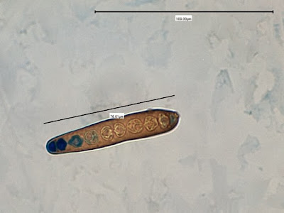
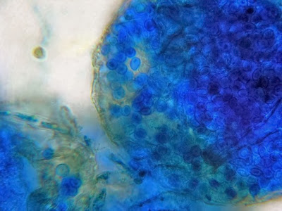
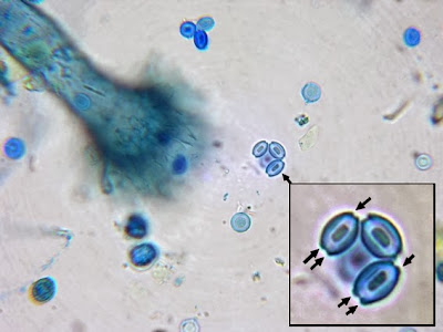
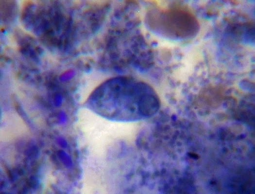
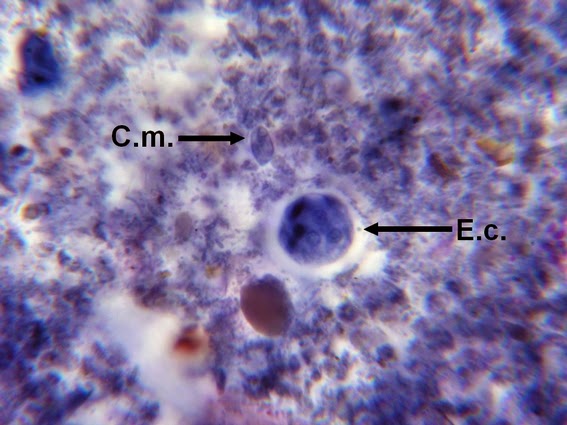
 Helicobacter heilmanii - Seen here in a gastric biopsy specimen (arrows).  The spiral appearance is much more evident in the enlarged insert.  The other large cells are gastric mucosal cells.  As we are only examining the biopsy for Helicobacter (pylori), which is a gram negative organism, there is no advantage to doing a full gram stain.  A small segment of the gastric mucosa, showing evidence of ulceration, was removed during an endoscopic procedure.  This tissue was mashed up onto a glass microscope slide and stained with carbol fuchsin.  (carbol fuchsin appears to stain these organism more intensely than gram safranin).  My experience has been that these gastric organisms seem to occur in 'pockets' in that you may search a slide for quite some time and not see a single organism, then suddenly, bang! several or even many in one area.  Pass over it and nothing once again.
(Nikon, Carbol Fuchsin Stain, X1000)

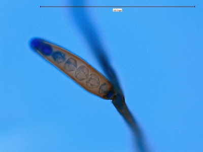
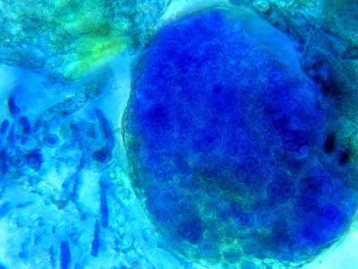
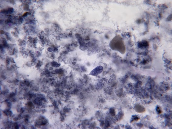
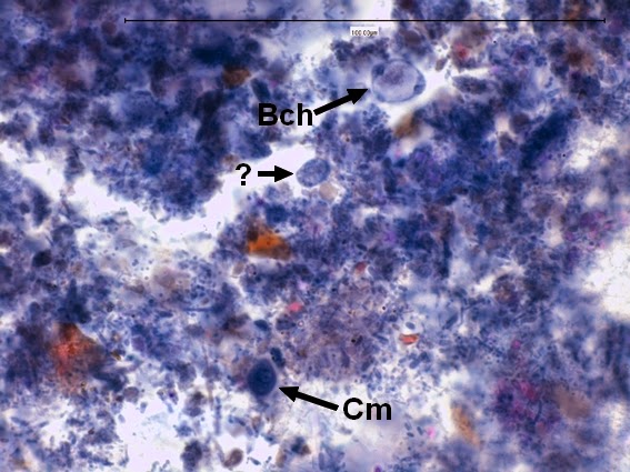
Heilicobacter heilmanii - from the same specimen as above.  The resolution is better in this photo with the tight wavey spirals (upwards of 8 in number) quite evident.  Organisms appear larger than above due to my cropping of the photograph.
 (Nikon, Carbol Fuchsin Stain, X1000) 

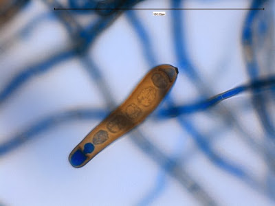
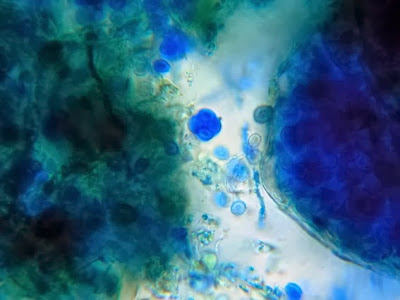
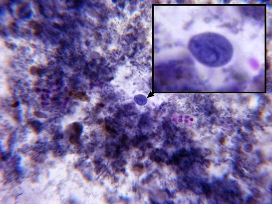
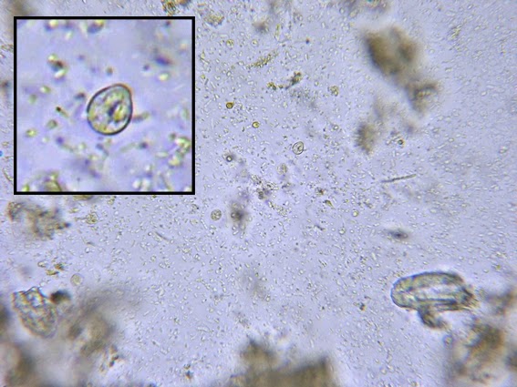
Helicobacter pylori - Compare the organism in the previous two photographs to Helicobacter pylori in this photo.  The arrow in the inset photo points to just one H.pylori organism showing the curved "S" shape or "gull-wing" shape of the bacillus.  Look carefully, the entire photo is teaming with H.pylori cells.
(Nikon, Carbol Fuchsin Stain, X1000)

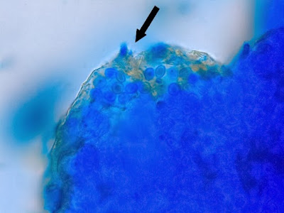
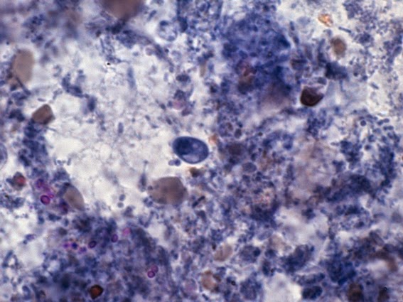
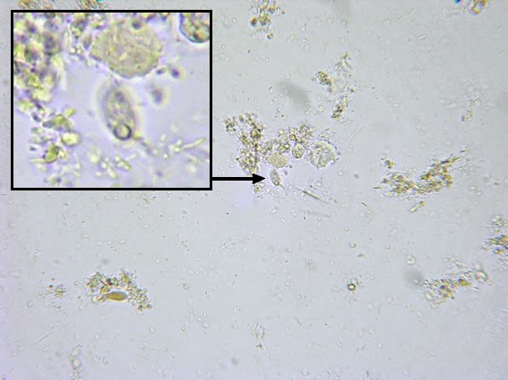
Campylobacter jejuni - Compare the Helicobacter species in the previous photographs to Campylobacter sp in the one immediately above.  Campylobacter also exhibits a curved or spiral (helical) morphology.  In fact, Helicobacter pylori was initially called Campylobacter pylori.  Here we can see a greater number of twists or spirals but there are not as many, nor are they as tightly wound as with H.heilmanii.  While C.jejuni is usually isolated from fecal specimens, this photo shows the organism in a blood culture.
(Nikon, Carbol Fuchsin Stain, X1000)

To read more about Campylobacter pylori, check out my Blog post specific to that organism by clicking here to redirect.


[i]Lancet 1987; 2:96
Dent, JC, McNulty CAM, Uff JC. Wilkinson, SP Gear MWL.
Spiral organisms in the gastric antrum.


[ii]Am J Gastroenterol. 1997 Aug;92(8):1310-5
Prospective, multivariate evaluation of CLOtest performance.
Weston AP, Campbell DR, Hassanein RS, Cherian R, Dixon A, McGregor DH.
Department of Veterans Affairs Medical Center, Kansas City, Missouri 64128-2226, USA


[iii]Rev Esp Enferm Dig. 1996 Mar;88(3):202-8.
C13 urea breath test in the diagnosis of Helicobacter pylori infection in the gastric mucosa. Validation of the method.
Pérez García JI, Pajares García JM, Jiménez Alonso I.


[iv]Eur J Gastroenterol Hepatol. 1999 Nov;11(11):1251-4.
The CLO test in the UK: inappropriate reading and missed results.
Prince MI, Osborne JS, Ingoe L, Jones DE, Cobden I, Barton JR.
University of Newcastle Regional School of Medicine, North Tyneside Hospital, North Shields, U


v Practical Gastroenterology, February 2006; 47-50
Clinical Significance of Helicobacter heilmanii Colonizing Human Gastric Antrum
Anup Hazra, Carlos Ricart and Januz J. Godyn
Dept of Pathology & Laboratory Medicine & Dentistry, New Jersey; Robert Wood Johnson Medical School, New Brunswick, New Jersey


vi Tzu Chi Med J, 2004; 16:  No1 59-62
Helicobacter heilmanii of the Stomach – A Case Report
Jeh-En Tzeng, Ying-Lung Lin, Yi-Tsui Chu, Sue-Mei Chung
Department of Pathology, Family Medicine, Buddhist Dalin Tzu Chi General Hospital, Chiayi, Taiwan
*   *   *

Arthrographis species

$
0
0


Arthrographis species (Mould)

Ecology:  Arthrographisis widespread, found in compost, decaying plant material and in the soil.

Pathology:  Arthrographisis generally considered to be a contaminant. Arthrographis kalrae has possibly been implicated in eye and central nervous system infections as well as onychomycosis (nail infections).  A case of ethmoid sinusitis has also been reported.  Arthrographis cuboidea* has been isolated from bronchial wash specimens however its pathogenicity remains unsubstantiated.  Other recognized species are A.lignicola, A.pinicola, A.sulphurea and A.alba however they have yet to be implicated as human pathogens.

*Recent molecular study appears to have reclassified this fungus, now known as Scytalidium cuboideum.  I will use the previous name throughout this post as I have found but a few references to this change.

Physiology:  Arthrographis cuboidea exhibits relatively fast growth at 25 – 30oC, maturing in about 3 to 5 days.    It grows equally well at 37oC at which temperature it may develop a pink to lavender pigment on prolonged incubation, however it fails to grow at 45oC  A.cuboidea possesses strong cellulolytic activity which can cause a pink coloured ‘spalting’ in many species of hardwoods.
 Arthrographis kalrae exhibits slow growth between 25 - 30oC, maturing within 1 to 3 weeks. Growth may be enhanced at 37oC, perhaps giving it some advantage as a human pathogen.   A.kalraeis capable of growing at 45oC which helps to distinguish it from A.cuboidea.  A.kalrae is resistant to cycloheximide.

Macroscopic Morphology:  (Species dependant)
A.cuboidea– rather fast growing as discussed above.  Colonies are generally described as cottony to granular.  Radial ridges or folds may develop.  Colouration is white to pale yellow with a yellow reverse.  A pink to lavender pigment may develop and diffuse into the media on prolonged incubation.
A.kalrae– is initially glabrous, smooth and yeast-like.  Colonies are slightly raised and become velvety or powdery as they develop.  Colour is described as a pale yellow (cream) to yellow-buff to tan.  The reverse is pale yellow to tan in colour.

 Arthrographis species - SAB, 72 hrs at 30oC

 Arthrographis species - Note no noticeable pink or lavender pigment on prolonged incubation.


Microscopic Morphology:  Both species produce hyaline, septate hyphae.  Arthrographis species develop conidiophores which differentiates this species fromGeotrichum& Scytalidium[i].  Conidiophores are generally short and can be branched or unbranched.  The arthroconidia which are produced from the conidiophores are smooth single celled and hyaline.  Arthroconidia produced from the conidiophores are rectangular to cylindrical in shape and are usually produced in chains.  With A.cuboidea, arthroconidia formed from undifferentiated hyphae are generally square or rectangular in shape.  A. kalrae may also produce lateral sessile (blastoconidia/aleuroconidia) which may be submerged in the agar and difficult to discern.  Intercalary arthroconidia may arise from undifferentiated hyphae and are generally longer and narrower than those produced from the conidiophores.  Arthroconidia separate by fission through double septa.

Note:  All photos which appear below were taken with the DMD-108 digital microscope.

 Arthrographis species - hyphae bearing conidiophores from which chains of arthroconidia extend.
(LPCB, X400)

Arthrographis species - as above but a closer look.  The larger or somewhat 'swollen' structures (arrows), point to the conidiophores.  Chains of arthroconidia are seen extending from the conidiophores.  (LPCB, X400+10, DMD-108)


Arthrographis species - another look at the septate hyphae, conidiophors and chains of conidia.  Note the 100 µm bar in upper right for scale.
(LPCB, X400)

 Arthrographis species - Branched hyphae with conidiophores and chains of arthroconidia
(LPCB, X400+10)

Arthrographis species - intercalary conidophores,  Chains of arthroconidia have collapsed around the tip.  No, this is not a mixed culture. (LPCB, X400)

 Arthrographis species - long chain of cylindrical or barrel shaped arthroconidia.  Smaller, narrower conidia (intercalary?) seen bunched may be those produced from an undifferentiated hyphae discussed above.  (LPCB, X 400+10)

Arthrographis species - Conidiophores bearing arthroconidia extending from hyphae.
(LPCB, 1000+10)

Arthrographis species - the two types of conidia as discussed above are shown in this photo.
(LPCB X400)

Arthrographis species - An older culture.  Septate hyphae with conidiophores bearing chains of arthroconidia.  (LPCB, X1000)

Arthrographis species - as above
(LPCB, X1000)
Arthrographis species - Again, an older culture - branched conidiophores extend from a hyphae with arthrospores collapsed around tip. 
(LPCB, X1000)

Arthrographis species - longer, narrower intercalary conidia extending from undifferentiated hyphae.
(LPCB, X1000)

Arthrographis species - as above (LPCB, X1000+10)

Arthrographis species - one feature I haven't found mentioned in any of my resource material is the small lateral structure at the tip of the conidiophore.  Any ideas?
 (LPCB, X1000)

Arthrographis species - the curious structure found at the tip of the conidiophore.
(LPCB, X1000)

Arthrographis species - One last view of the curious lateral structure found at the tip of the conidophores of this particular species.  I have not found mention of this structure in any of my resource material.  (LPCB, X1000+10)


Note:  I won’t venture a guess at what specific species I’ve isolated here.  Growth was quite rapid and produced a pale yellow pigment favouring C.cuboidea.  I did not attempt temperature studies.  While the species survived on Dermasil™ media containing cycloheximide, growth was weak and stunted.  Only molecular studies would provide the definitive identification.


[i] Recent molecular study appears to have reclassified this fungus, now known as Scytalidium cuboideum.  I have not found reference to this reclassification nor how the presence or absence of conidiophores is reconciled.

Trichothecium roseum

$
0
0


Trichothecium roseum  (Fungus)

Ecology: Trichothecium species are cosmopolitan fungi (found just about everywhere) and is a common saprobe (growing on decaying vegetation).  In particular, it has been isolated from withering fleshy fruits such as peaches, plums and nectarines.  Trichothecium is responsible for ‘pink rot’ of apples.

Macroscopic Morphology:  Trichothecium is a rapidly growing fungus which is fully mature in 3 to 4 days.  Colonies are described as being flat, suede-like to powdery, which start off white but quickly develop a light pink to peach colour which may reach a darker salmon colour on continued incubation.  The reverse is a rather non-descript light or pale colour.  Pictured below are colonies grown at 30oC on Sabouraud Dextrose Media (SAB or SDA).  Trichotheciumfails to grow at 37oC making it an unlikely candidate for human pathogenicity.

 
 Trichothecium roseum on SAB agar, 72 hrs, 30oC (Nikon)
It was rather difficult to capture (and correct for) the exact colour hue under harsh labor fluorescent laboratory lighting within the biological laminar flow hood.  The colour descriptions range from a light pink to peachy to salmon or even orange on extended incubation.

Trichothecium roseum on SAB after 14 days incubation at 30oC (Nikon)

Microscopic Morphology:  Hyphae produced by Trichothecium are septate and hyaline (clear, not pigmented).  The long, thin and erect conidiophores are indistinguishable from the vegetative hyphae and may exhibit septation near their base of attachment.   Trichothecium roseum produces rather thin walled, two-celled conidia (16-20µm X 8-12µm) which are pyriform or clavate in shape.  Basipetal growth has the newest cell developing below the previous one (The youngest cells are at the base while the oldest are at the apex.)  This growth produces a sympoidal pattern seen as zigzag or alternating conidia extending from the conidiophore on opposite sides.  Free conidia have a truncated basal scar usually obliquely offset, indicating their former point of attachment.

Note: All photos which appear below were taken with the Leica DMD-108 digital microscope.

 Trichothecium roseum growing from the surface edge of agar (bottom of photo).  Fine hyphae and conidiophores bearing conidia are seen (7 days 250X LPCB)

Trichothecium roseum -conidiophores are seen extending along the lengths of hyphae.  Conidia are seen clumped at the apex of the hyphae.  Branching of conidiophores is rare if it occurs at all.
(250x. LPCB)

Trichothecium roseum - conidiophores with early production of two-celled conidia are seen extending from a hyphal element just out of focus below.  A number of two-celled conidia are seen free of the conidiophore.  A truncated basal scare can be seen which may may be somewhat offset from center (arrow), due to the sympodial pattern of growth.
(400X, LPCB)

Trichothecium roseum - again, conidiophores are seen extending from the hyphae (slightly out of focus at top).  Cells appear 'clustered' around the apex of the conidiophore as the growth extends.  Note 100µm bar at top right of this and previous photo.
(LPCB, 400X)

Trichothecium roseum - conidiophores extending from hyphae with pyrimidal or clavate shaped (pear shaped) conidia extending to the apex.  Again, 100µm bar appears on this and various other photos for scale.  (400X, LPCB)

Trichothecium roseum - somewhat thick walled, two-celled clavate conidia are seen at the apex of the conidiophore extending upwards into the focal plane of the camera.  The conidium at the top of the group appears contorted (twisted) at the bottom where it is attached to the conidiophore.  When released, a truncated basal scar will be present at this attachment point and it will be somewhat offset from the centerline of the conidium. (1000X, LPCB)

Trichothecium roseum - once again the clavate shaped conidia are seen attached along the conidiophore.  Here you can see the sympodial growth pattern which produces conidia in an alternating or zigzag pattern along the conidiophore.  The youngest cells are at the bottom with the most mature at the apex (top) of the cluster.  (1000X, LPCB)

Trichothecium roseum - free conidia showing the pyramidal, clavate (club shaped) or perhaps pear shape characteristic of this fungus.  The cell wall is rather thin to moderately thickened and the central division is visible in most cells above.  Again, the basal scar appears at the previous point of attachment and may be somewhat offset from the center line of the conidium.
(1000X, LPCB)

Trichothecium roseum - clavate two-celled conidia seen attached to conidiophore
(1000+10X, LPCB)

Trichothecium roseum - one more photo just for the heck of it.  Septate hyphae can be seen.  Sympodial attachment of cells can be seen with the two cells near the center of the photo.
(400X, 14 days, LPCB) 

Trichothecium roseum -Computer wallpaper (1024X768)
 
Pathogenicity:  Trichothecium species are generally clinical laboratory contaminants.  No human or animal infections have been reported.

Differentiation:  Trichothecium roseum may initially be confused with Microsporum nanum as this fungus produces a similar light pink to buff coloration.  M.nanum, however, exhibits a reddish-brown pigment on reverse in contrast to the pale reverse of T.roseum.   The conidia produced by M.nanum are also two celled however they are sessile (attached directly to undifferentiated conidiophores) or on short stalks.  Finally, Microsporum nanum has the ability to perforate hair cells and is not inhibited by the cycloheximide in Mycosel agar.
*   *   *

Trichophyton mentagrophytes Complex

$
0
0


Trichophyton mentagrophytes Complex(Fungus, Dermatophyte)

Ecology:  T.mentagrophytes is recognized as having two variants.  Anthropophilic isolates prefer man to animals while the zoophilic isolates primarily infect animals.  Small rodents appear to be the primary reservoir for the animal variety.  Trichophyton mentagrophytes is a cosmopolitan fungus (found everywhere).

Macroscopic Morphology:  T.mentagrophytes exhibits moderately rapid growth and matures within 6 – 10 days.  Sources have previously described anthropophilic isolates having a downy, powdery or even fluffy texture while zoophilic isolated were more granular in appearance.  Colonies may vary in colour from white to cream or yellowish.  The reverse also can vary from yellow to reddish brown to brown or ochre, depending on isolate and medium.

 Trichophyton mentagrophytes -5 days growth on SAB at 30oC

 Trichophyton mentagrophytes - 14 days growth on SAB at 30oC


Microscopic Morphology:  Trichophyton mentagrophytes produces septate hyphae from which branched conidiophores extend.  Sessile (not on stalk) microconidia are produced in rather dense, grape-like clusters on the conidiophores.  The microconidia (~2µm to 4µm) are spherical to pyriform in shape.  Macroconidia (20-50µm to 6-8µm) are cigar to club shaped and may show exhibit some distortion.  Macroconidia have a smooth exterior and are thin walled, usually have between 3 to 8 cells dividing the interior.  Macroconidia may be found more readily in younger cultures.  Production of both micro & macro conidia may vary with the isolate.  Coiled or spiral hyphae may be present and in some strains, structures described as nodular bodies or chlamydospores may be present.

Note: All photos which follow were taken with the DMD-108 digital microscope. 

T.mentagrophytes showing sessile microconidia along septate hyphae.
Note 100µm bar in upper right of this and several other photos. 
(400x, LPCB)

Trichophyton mentagrophytes - branched conidiophores bearing spherical conidia in clusters seen extending from septate hyphae.  (400x, LPCB)

T.mentagrophytes - another view as above.  A macroconidium can be seen near the lower center of the photo (400x, LPCB)

T.metagrophytes - a closer look at the branched condiophores bearing clusters of spherical microconidia.  Septations are visible in the hyphae and conidiophores.
(1000x, LPCB)

T.mentagrophytes -another look as in the previous photo.
(1000x, LPCB)

T.mentagrophytes - A 7-celled macroconidium.  Macroconidia are typically described as being cigar shaped or club shaped.
(400x, LPCB)

T.mentagrophytes - another 7-celled macroconidium with dimensions (inset)
(400+10x, LPCB)

T.mentagrophytes -an macroconidium which shows slight distortion (sides are not straight). Numerous spherical microconidia in lower right.
(1000x, LPCB)

T.mentagrophytes - a solitary cigar shaped macroconidium showing seven internal cells.  Walls are rather thin and the exterior is smooth.
(1000+10x, LPCB)

T.mentagrophytes - just by chance all this, and the previous macroconida all contain seven cells.  I found that young cultures (~3 days) produced the most macroconidia which seemed to diminish with additional incubation.  That said, the macroconidium pictured here is from a 6 day old slide culture.  It should always be kept in mind that structures may appear, disappear, develop or change with length of incubation.  It may be advisable to make several side cultures and harvest them at different time periods to observe development of structures.
(1000+10x, LPCB)

T.mentagrophytes - a couple of macroconidia are seen in this photo as well as clusters of microconidia.  A spiral hyphal element is seen in the upper center of the photo.
 (400+10x, LPCB)

T.mentagrophytes - a spiral hyphae is seen seen in the center left of this photo as it overlaps a macroconidium.  Microconidia throughout the photo.
(1000x, LPCB)

 T.mentagrophytes - more examples of spiral hyphae typical to T.mentagrophytes seen in this photo.
(400+10x, LPCB, 8 days incubation)

T.mentagrophytes - one last photo showing what is described as a nodular body or chlamydospore.  Clusters of microconidia seen in center-left.
(1000+10x, LPCB, 10 days incubation)

Physiological Tests:  a number of classical tests can be employed to speciate Trichophytonspecies.
·         Urease test: Positive
·         BCP-Milk Solids Glucose: Alkaline reaction
·         Hair perforation test: Positive
·         Growth at 37oC: Excellent
·         Growth factor requirement*: None
*a variety of Trichophytontubed agars are commercially available containing specific growth supplements (eg.inositol, thiamine, nicotinic acid, histidine).  The pattern or degree of growth in each can assist the speciation of Trichophyton.

Pathogenicity:  The anthropophilic strains are usually associated with chronic infections of glabrous skin, scalp, beard, nails and feet.

It is currently recommended* that Trichophyton mentagrophytes be reported as ‘Trichophyton mentagrophytes complex’ which also includes the former Trichophyton krajdenii.

*Best Practice by QMP-LS, external quality assessment agency.

*   *   *

Phoma glomerata

$
0
0


Phomaglomerata(fungus)

Ecology: Phomais yet another ubiquitous, cosmopolitan fungus which is commonly found in soils.  As a known plant pathogen, it may also be recovered from infected plant material.  Phoma species may be found in the laboratory as an environmental contaminant.

Macroscopic Morphology:  Phoma is a rapidly growing fungus which usually reaches maturity within five days.  The full development of picnidia may take somewhat longer.  Colonies have are usually described as having velvety texture though some sources add they may appear powdery to even woolly.  Colonies are usually described as brown to olivaceous and even grey in colour.  The reverse is brown to dark brown to black with some species producing a diffusible reddish-brown pigment.

 Phoma glomerata - 5 days on SAB at 30oC

 Phoma glomerata -Same colony as above at 14 days incubation at 30oC
(Lighting differs from the photo above - difficult to control while trying to capture the true hue)


Microscopic Morphology:   Phomaglomerata produces sub-hyaline to hyaline (dark pigmented/brown), septate hyphae.   Phomaproduces rather large (~60 µm to 400 µm) pyriform to globose shaped pycnidia[i].  Phialides line the interior of each pycnidium, which produce single celled (rarely two-celled) hyaline (clear) to pale brown, ovoid to ellipsoidal (5-10 µm by 2.5 to 3.0 µm) conidia.  The conidia have been described as bi-guttulate (containing 2 oil droplets).  Mature conidia are released from the interior of the pycnidium through an ostiole (pore or opening).  Phoma glomerata also produces chlamydospores (chamydoconidia) in branched or un-branched chains.  The chlamydospores may show both longitudinal and transverse septations (muriform) as is commonly seen in the genus Alternaria

Note:  All photographs which appear below were taken with the Leica DMD-108 digital microscope.

 Phoma glomerata - edge of slide culture showing hyphae with the development of pycnidia.
(LPCB, 250X)

Phoma glomerata - pigmented chlamydiospores (chlamydioconidia) extending from hyphae.
(LPCB, 400X, 12 days)

Phoma glomerata - as above.  Longitudinal and transverse (muriform) septations are visible within the chlamydospores at this magnification.  Free conidia are seen throughout photo.
(LPCB, 400X, 12 days)

Phoma glomerata - muriform chlamydospores.
(LPCB, 400+10X, 12 days)

Phoma glomerata - Pigmented chlamydospores.  Both longitudinal and horizontal septations are easily seen in the chlamydospore in this photograph (arrow)
(LPCB, 400+10X, 12 days)

 Phoma glomerata - Pycnidia seen as darkly pigmented bodies.
(LPCB, 250+10X)

Phoma glomerata - Three pycnidia showing their brown pigmentation in a LPCB preparation.
(LPCB, 400X)

Phoma glomerata - not the greatest photo example, but I like it.  Pycnidium showing a massive release of conidia from within.
(LPCB, 400+10X, 12 days)

Phoma glomerata - as above.  LPCB prep, however the cells are so dense that the stain hasn't penetrated throughout the preparation.
(LPCB, 400X)

Phoma glomerata - a pyriform shaped pycnidium is seen with the ostiole at center left.  Interior structure of the pycnidium can be somewhat visualized through the wall. Conidiophores line the interior wall of the pycnidium where conidia are produced.  Free conida can be seen throughout the photo.  Pigmented septate hyphae are also seen in the background.
(LPCB, 400+10X)

Phoma glomerata - three, possibly four pycnidia.  Septations are clearly visible in the hyphae at the center of the photograph.
(LPCB, 400X)

Phoma glomerata - possible small ostiole seen (arrow)
(LPCB, 400+10X, 5 days)

Phoma glomerata - Pycnidium with prominent ostiole visible, trailing conidia.
(LPCB, 400+10X, 12 days)

Phoma glomerata (LPCB, 400X)

Pathogenicity:  Phomaspecies are known to be pathogenic to some plants however both human and animal infection is infrequent.  Phomaglomerata has been implicated in a few documented cases of phaeohyphomycosis.   It has also been reportedly isolated from an ulcerated human cornea.

Caution:  The pycnidia produced by Phoma species should not be confused with the perethecia  suchas those of Chaetomium species or the cleistothecia of Pseudallescheria boydii.


[i] An often flask shaped conidiomata of fungal tissue which is lined on the inside with conidiophoresAn asexual fruiting body.

*   *   *

Aspergillus versicolor

$
0
0


Aspergillus versicolor(mould, fungus)

Ecology:

Widespread distribution with ability to grow in temperate and colder regions which may restrict the growth of many other Aspergillus species.    Commonly found in soils and may be isolated from many foods, especially spices, dried cereals and nuts.  Frequently found in buildings with humidity & ventilation problems.

Pathogenicity:

Has been implicated as the causative agent of a variety of human mycoses including onychomycosis, pulmonary disease and disseminated infection.  Association with disease is poorly documented which leaves doubt to its significance.

Macroscopic Morphology:
  • A.veriscolorhas a slow to moderate rate of growth, maturing in about 3 to 7 days.

  • Expansion of colony is rather slow.

  • Growth is restricted at 35ºC.

  • Colony texture is suede-like, often with radial grooves.

  • Colonies can develop a variety of colours (hence the name – “versicolored”).

  • Colour ranges from pale green, greenish-beige, grey green, pinkish-green, salmon-green to dark green with shades of mauve or turquoise. (colour development is media dependant)
  • Exudate when present may be pint to reddish-brown.

  • Reverse is uncoloured to a reddish-brown or reddish-purple.

Aspergillus versicolor SAB 14 days 30ºC


Aspergillus verisicolor ~12 days - This photo taken from a previous isolate better shows the variability of the color produced by A.versicolor.  This species is capable of producing a variety of colors, hence the name - verisi-colored.

Microscopic Morphology:

Smooth conidiophores (~ 200 – 500 µm X 4 -7 µm) extend from septate hyphae.

Conidial heads support vesicles (9-16 µm dia) which are biseriate with metulae about the same size, or slightly shorter than the phialides.

The conidiogenous cells (metulae & phialides) loosely cover half to the entire vesicle.

Diminutive conidial heads can form which can resemble penicilli (Mimics Penicillium species structure).

Conidia (2-0 – 3.5 µm dia) are globose (round) and the walls usually have a slightly roughened appearance.

Globose hülle cells on occasion are present in some isolates.

 A.verisicolor -Edge of a slide culture at 48 hours of incubation.
 (LPCB, 250X, DMD-108)

A.verisicolor - fruiting structures
(LPCB, 400X, DMD-108) 

Ditto

A.verisicolor -conidiophore -Stipe with vesicle at apex which is covered with metulae & phialides (Biseriate) that produce the conidia.  (LPCB, 400X, DMD-108)

A.veriscolor - just a closer look at the photo above adding 10X digital magnification to the optical.
(LPCB, 400+10X, DMD-108)

A.versicolor -I like photographs.  Another photo of the fruiting structure of A.versicolor.  Note the 'reduced' structures at the left of the photo.  More on those to come.
(LPCB, 400X, DMD-108)

A.verisicolor - yet another view
(LPCB, 400X, DMD-108)

A.versicolor -Biseriate structure (metulae & phialides) but difficult to tell in this photo.  Abundant conida seen chaining from the phialides.
(LPCB, 1000X, DMD-108)

A.versicolor -again difficult to see the biseriate structure at this resolution.  Image structures and colours seem to "bleed" together with the DMD-108 digital microscope.  Reducing saturation of the image did not help.
(LPCB, 1000+10X, DMD-108)

A.versicolor - Biseriate structure, although not tremendously clear, can be visualized in places in this photo. (LPCB, 1000+10X, DMD-108)

A.versicolor - somewhat of a messy photo, lacking clear structures, but I like it, so it is here!
(LPCB, 1000+10X, DMD-108)

A.versicolor - one characteristic of A.versicolor which aids in its identification is that it produced both fruiting structures typical of Aspergillus genus, the vesicle covered with conidiogenous cells (A), but also a reduced structure which can resemble penicilli  (B). (ie. somewhat looks like a Penicillium fungus.)  (LPCB, 400X, DMD-108)

A.versicolor - another photo where both structures (as described above) are seen in the same field.
(LPCB, 400X, DMD-108)

A.verisicolor - as above but clearer view of the reduced structure seen in the inset.
(LPCB, 400X, DMD-108)

A.versicolor - reduced penicilli structure.
(LPCB, 400X, DMD-108)

A.versicolor - another view of the reduced penicilli structure.  The variable color and the presence of these reduced structures are distinctive features of A.versicolor, thereby aiding in identification.
(LPCB, 400X, DMD-108)

A.versicolor - reduced penicilli structure.  No vesicle is present.
(LPCB, 1000+10X, DMD-108)

A.versicolor - this photo is taken at the edge of a slide culture after 48 hours incubation.  Numerous reduced forms appear to be present.  Background of photo is due to agar from the slide culture preparation remaining attached to the coverslip.
(LPCB, 400X, DMD-108)

A.versicolor - Conidia (2-0 – 3.5 µm dia) are globose (round) and the walls usually have a slightly roughened appearance.
(LPCB, 1000+10X, DMD-108)


Notes:

Caution – A.versicolor’s fruiting structure may at first observation be confused with A.sydowii.   Colonial appearance differs substantially where A.sydowii has a distinctive blue-green colour in comparison to A.versicolor’svariable but dominant light-green shades.


*   *   *

Entamoeba polecki

$
0
0


Entamoeba polecki

This amoeba most likely has worldwide distribution and can be found as an intestinal parasite in pigs and monkeys.  It is generally considered to be non-pathogenic for humans but has occasionally been recovered from patients with loose stools and intestinal discomfort.  Several sources have stated that in Papua New Guinea, E.polecki has been recovered as the most common intestinal parasite from the inhabitants.  Living closely with these animals my increase the chances of carrying the parasite.  E.polecki makes its home in the large intestine of man.

Trophozoites:
E.poleckitrophs have an average size of 12 µm to 18 µm but can range from 10 µm to as large as 25 µm.  Its cytoplasm can be vacuolated and may contain ingested bacteria and yeast.  The size and cytoplasmic appearance may cause it to be confused with Entamoeba coli.  The karyosome may not be visible which has the nucleus appear empty.  When present, the karyosome is usually minute and centrally located, or nearly so.  It however may occasionally appear diffuse.  Peripheral chromatin can vary from being fine and evenly distributed as in E.histolytica, or coarse and irregularly distributed as in E.coli.  This variability may make diagnosis a challenge.

Cysts:
E.poleckicysts are about 9 µm to 15 µm but can be as large as 24 µm and usually contain only one nucleus (usually E.histolyticahas 4, E.coli has 8).  It has rarely been reported as being binucleate.  The karyosome again can be minute can compact or it may be larger and diffuse – it may be centrally located within the nucleus or somewhat eccentric.  Peripheral chromatin may be delicate to coarse but is evenly distributed on the nuclear membrane.  Chromatoidal material is usually abundant but highly variable in shape and size.  It may be present as larger rods with rounded or splintered ends and they may be arranged parallel to each other in the cyst.  Unique to these cysts may be an inclusion body of variable size.  It stains a monochromatic grayish-purple with Iron hematoxylin stain or greenish with the trichrome stain.  In an iodine stained preparation it usually appears light brown in contrast to the intensely dark brown staining of glycogen vacuoles.  The nature of these inclusions is not known but does not appear to be glycogen.

Differentiation:
Differentiating the E.polecki trophozoites from E.histolytica and E.coli may be difficult as the size ranges overlap and both the cytoplasmic and nuclear appearance can mimic both.  If examining a specimen that has amoeba showing characteristic of both E.histolytica& E.coli, the presence of E.polecki should be considered. Chromatoid bodies are usually are more numerous and show greater pleomorphism in E.polecki than E.histolytica.  E.polecki cytoplasm often stains very darkly making it difficult to see the nucleus, inclusion mass and chromatoid bodies in the same plane of focus

Note: The photographs which follow were of E.poleckifound in a fecal specimen obtained from a patient with gastrointestinal discomfort.  Only trophozoites were present in the sample.  I’m rather unhappy with the quality of the photos I took and present here.  The features lack resolution and the amoeba appears darker in the photos than they did when viewed through the light microscope.  The features appear to be over saturated in colour and “bleed” together, obscuring details.  I was unable to correct for this satisfactorily with photo software post exposure.  This specimen also contained Entamobea hartmanni trophs.  Identification was confirmed by our provincial public health laboratory. Here they are for what they’re worth.

All photos below were taken using the Nikon 'Coolpix' camera at 1000X magnification and are from an Iron-Hematoxylin stained preparation.

E.polecki trophozoite exhibiting a coarse cytoplasm.  Peripheral chromatin in the nucleus is rather evenly distributed.  Karyosome is central but not distinct.

E.polecki trophozoite showing rather evenly distributed peripheral chromatin in the nucleus and as above the karyosome is rather diffuse and slightly eccentric.

E.polecki trophozoite showing a rather coarse cytoplasm with ingested material (bacteria).  The karyosome shows even, dense peripheral chromatin but the karyosome appears to be absent in all planes of focus. Above the E.hartmanni troph may be a somewhat distorted Blastocystis hominis.

E.polecki troph as above.  "Dirty" cytoplasm, even dense peripheral chromatin in the nucleus with a somewhat diffuse karyosome, slightly eccentric.

This specimen contained both E.polecki trophs and E.hartmanni trophs.  Note the size difference.

E.hartmanni troph and a second cell with too little detail to identify confidently.

E.policki troph (inset left) to demonstrate the size difference between it and the E.hartmanni troph.

*  *  *


Trichophyton terrestre

$
0
0
Trichphyton terrestre (Fungus/Mould)

Ecology:
Trichphyton terrestre is a cosmopolitan (found everywhere), geophilic (soil loving) fungus.
It may also be recovered as a saprobe (living on dead organic matter) on the fur of small animals, presumably picked up from soils.

Pathology:
Trichophyton terrestre fails to grow once 35ºC to 37ºC is reached
As it fails to grow at human body temperature, there have been no reports of human infection by this fungus, nor has it been implicated in animal infections.
T.terrestre may occasionally be encountered as a laboratory contaminant.
Correct identification is important so as to not confuse it with pathogenic dermatophytes.

Macroscopic Morphology:
  • Trichophyton terrestre exhibits moderate growth at 25ºC, maturing in about 8 days.
  • Colonies expanded in diameter rather slowly.
  • Colonies were off-white to light in colour.
  • Reverse appeared yellowish to ochraceous, or even slightly reddish in colour.
  • Texture was felty to powdery
The isolate presented here developed a pale to golden yellow exudate on prolonged incubation which was not reported in other sources.


 Trichophyton terrestre on SAB, 15 days incubation at 30˚C

Trichophyton terrestre on SAB,  25 days incubation at 30˚C


Microscopic Morphology:
  • Trichphyton terrestre produces hyaline (clear, non-pigmented), septate hyphae.
  • Microconidia (4 – 7 µm by 1 -5 µm) are tear-drop to slightly club-shaped.
  • They are borne directly from the vegetative hyphae or are found on pedicles (stalk).
  • Macroconidia (4 – 5 µm by 8 – 50 µm) have smooth, thin walls and usually contain between 2 to 6 cells or divisions internally.
  • Macroconidia are cylindrical (parallel sides) or slightly clavate (club) shaped.
There may not be an obvious distinction between what may be called micro or macro conidia.  (ie. The two are not clearly differentiated.)
Free micro (& macro conidia) exhibit a truncate base or basal scar at what was their point of attachment.

Trichophyton terrestre - Adhesive tape preparation, 250X, LPCB (Nikon)

Trichophyton terrestre - Branching with development of Macro & Micro Conidia.
 LPCB 400X (DMD-108)

Trichophyton terrestre -as above
 LPCB 400X (DMD-108)

 Trichophyton terrestre - free macro & micro conidia
400+10X, LPCB (DMD-108)

Trichophyton terrestre - Conidia stain darker with the LPCB than the hyphae usually do
1000X, LPCB (DMD-108)

Trichophyton terrestre - Here again, the conidia at the tips of the hyphae (conidiophore) can be seen as staining a darker blue than the hyphae themselves.  Measurements shown (inset) are for the conidia and hyphe.  I regret that I didn't just measure the length of the conidia alone.
1000X, LPCB (DMD-108)

Trichophyton terrestre -extensive branching at near right angles.
1000X, LPCB (DMD-108)

Trichophyton terrestre -more of the same.  Darker blue conidia are seen at the end of the hyphae, branching at near right angles.
1000X, LPCB (DMD-108)

Trichophyton terrestre - divisions can be seen in some of the developing conidia (macroconidium)
1000X, LPCB (DMD-108)

Trichophyton terrestre -conidia staining a darker blue with the LPCB stain.  Divisions can be seen in the conidia.  The one on the lower left of the photo clearly has two.
1000X, LPCB (DMD-108)

Trichophyton terrestre -free macroconidium (4 septations or 5 compartments)
1000X, LPCB Nikon (appears larger due to cropping of photo)

Physiological Characteristics:
  • Hair perforation test is POSITIVE
  • Urease is POSITIVE
  • BCPCG Media reaction is POSITIVE
  • No growth at 35ºC to 37ºC
Trichophyton Agars:
Good growth on all Trichophyton agars.  No special growth requirements are required for growth.
Caution: on early growth, the fungus may somewhat resemble a Chrysosporiumspecies.  Chrysosporium’s conidia generally do not exceed two cells in length.

*  *  *

Exserohilum rostratum

$
0
0


Exserohilum rostratum (Mould)

Ecology:
Exserohilumspecies are dematiaceous fungi (ie black mould), widely distributed in nature.  They are cosmopolitan, commonly found on many plants and grasses and can also be isolated from soils and water.

Pathogenicity:
Exserohilum species are unlikely human pathogens.  Exserohilum species have been implicated in phaeohyphomycosis[i]. Most commonly they are isolated from nasal sinuses (sinusitis) and the eye (keratitis) after a scratch or traumatic injury.

In 2012, Exserohilum rostratum was implicated as the primary pathogen isolated from injectable methylprednisolone. Numerous patients receiving steroid therapy primarily for degenerative lumbar-disk and joint disease in the U.S., developed meningitis after injection. While normally not invasive, the fungus will cause illness when directly injected into the body. While sources vary in statistics, injectable methylprednisolone contaminated with Exserohilum rostratum appears to have been responsible for sickening upwards of 700 people, 30 of which died.  Many others continue to have ongoing neurological problems associated the infection or subsequent therapy.

Macroscopic Morphology:
E.rostratumexhibits rapid growth and matures within 4 to 5 days.
Surface growth is grey in colour, quickly darkening with the production of melanin, eventually developing shades from olive to brown to black.
The texture is woolly or cottony in appearance.

Exserohilum rotratum SAB 30oC, 72 Hours

Exserohilum rostratum SAB 30oC, 1 Week


Microscopic Morphology:
Hyphae are septate and darken with the development of melanin.  Conidiophores are rather long (up to 200 - 230 µm in length, 5 – 8 µm wide) and are also septate.  Conidiophores exhibit sympodial geniculate growth, where conidia are produced at bends (geniculate) as the conidiophore extends.  This gives the conidiophore a knobby, zigzag appearance where the conidia attach.  The mature conidia (ave. 14 X 80 µm or greater) are straight to slightly curved and are fusiform or ellipsoidal in shape with rather smooth walls.  Conidia are compartmentalized with between 7 to 11 septa and has a distinctive protruding dark hilum (scar) at the base where once attached.
More specifically, the conidia are ‘poroconidia’, a distinction where the conidia are produced through the extrusion or extension of the inner walls of the conidiogenous cells through a pore or channel.

 Exserohilum rostratum -First look.  Free conidia.  Tape mount at 250X (LPCB, DMD-108)

Exserohilum rostratum -Conidia attached to septate hyphae (400X, LPCB, Nikon)

Exserohilum rostratum - Large, compartmentalized conida which are somewhat fusiform in appearance and may appear slightly bent or curved.  (400X, LPCB, DMD-108)

Exserohilum rostratum - as above.  Showing variation in size and shape of conidia.  Brown pigmentation due to the production and accumulation of melanin. (400X, LPCB, DMD-108)

Exserohilum rostratum - Conidia attached to conidiophore.
(400X, LPCB, DMD-108)

 Exserohilum rostratum - as above, conidia attached to conidophore seen extending through the camera's plane of focus.  (400X, LPCB, DMD-108)

Exserohilum rostratum - a closer look at the septate conidiophore and the attachment of the conidia to the conidiophore.  Note the bent or zig-zag location on the conidiophore at the point of conidial attachment.  This appearance us referred to as geniculate growth.  (400X, LPCB, Nikon)

Exserohilum rostratum - Pigmented, fusiform shaped conidia, usually containing between7 to 11 internal septa. Note the prominent projection or hilum (arrow) which remains at the point of the attachment to the conidiophore. (400+10X, LPCB, DMD-108)

Exserohilum roatratum - single conidium with hilum visble on right side.  Length reads 76.01
µm. (400+10X, LPCB, DMD-108)

Exserohilum rostratum - another view of the geniculate growth / attachment of the conidia to the conidiophore. (400X, LPCB, DMD-108)

Exserohilum rostratum - Single conidium attached to conidiophore.
(1000X, LPCB, DMD-108)

Exserohilum rostratum - okay, I like photos!  Another loose conidium which is slightly bent.  This one has seven, possibly eight compartments.  The conidium is smooth walled and again, the hilum is clearly visible at one end.  (1000X, LPCB, DMD-108)

Exserohilum rostratum - More photos, just for the beauty of this organism.
(1000X, LPCB, DMD-108)

Exserohilum rostratum

Exserohilum rostatum - geniculate (zig-zag) conidiophore after the conidia have dispersed.
(400X, LPCB, DMD-108)
 Caution:
Exserohilum species may be confused with  Bipolaris and Drechslera species however Exserohilum has the protuberant hilum.


[i] A particular presentation of a fungal infection in tissue caused by certain dematiaceous fungi.  This presentation may be an initial clue as to the particular fungus responsible for the infection.  'Google' the term for a better definition.

*   *   *

Eurotium herbariorum (Aspergillus glaucus)

$
0
0


Eurotium herbariorum(Aspergillus glaucus) Mould

Note:  This fungus has both a sexual and asexual means of reproduction.  When both are present, the sexual stage (teleomorph) name takes precedence over the asexual (allomorph) name.  Applied here, Eurotium herbariorum take precedence over Aspergillus glaucus.  However,  I will generally refer to the fungus here as Aspergillus glaucus as most reverences still continue to do so.

Ecology: 
Aspergillus glaucus is a cosmopolitan fungus (worldwide distribution) and while it prefers drier environments, it can be isolated from soils, house dust, plants & dried foods.  A.glaucus is also osmophilic, meaning it can grow, and perhaps prefers to grow in environments containing a higher sugar concentration.  Growth is restricted or limited at 35˚C which may account for its limited pathogenicity.

Pathology:  
A.glaucus is not very invasive and is rarely encountered in the clinical laboratory.  It has been implicated as a cause of ocular (eye) infections, particularly after some traumatic injury.  Cerebral, orofacial, cardiovascular and pulmonary infections are rare but have been reported.  May also cause sinusitis (nasal) and otitis (ear) infections.  May be considered an opportunistic fungus particularly with immunocompromised patients.

Macroscopic Morphology:
Growth is slow to moderate, maturing in about 7 to 21 days.  Colony size expands rather slowly.
Colony colouration is media dependent but is described as a dull to deep green to a greyish turquoise, with yellow to orange areas where cleistothecia are being produced.  The reverse is pale yellow to yellow.  The isolate presented below failed to grow at 30˚C but grew well at ambient room temperature (~20˚C).
 Eurotium herbariorum (Aspergillus glaucus) -SAB + ~20% Sucrose, Room Temperature, 14 Days
(this particular isolate failed to grow at 30˚C)

Note on Culture:  Our medical laboratory purchases all media from a commercial supplier and in these financially challenging times we stock only what can be clinically justified.  We have no media room where we can concoct media from basic ingredients.  Sabouraud Dextrose media has a sugar content (dextrose) of 4% which would favour the production of asexual conidia while a high osmotic concentration (~20%) would enhance cleistothecia production.  In order to enhance growth and increase the production of cleistothecia, I obtained a couple of tubes of Sabouraud Dextrose (slants) and melted them down in a boiling waterbath.  I did a rough 'back of an envelope' calculation of how much sugar I would have to add to bring the concentration up to about 20%.  With no lab supply of dextrose to add, I went down to our hospital 'Tim Horton' coffee shop (a Canadian franchise) and obtained a packet of sucrose.  Adding this to the melted SAB agar, I brought the total sugar concentration up to about 20% (glucose + sucrose) to raise the osmotic concentration.  I sacrificed a sterile media petrie dish contents (as we also do not stock sterile empty petrie dishes) and poured in my own concoction of "Tim Horton's Cleistothecia Enhancing Media", or THCEM for short.  For this reason you will see the media identification on the reverse plate blurred out. With no autoclave on site, I hoped that the boiled contents remained sterile and the high osmotic concentration would discourage other organisms from growing.  The THCEM media worked extremely well!  The photographs which follow are from this agar media.

Microscopic Morphology:
Hyphae are septate and hyaline.
Teleomorph– Sexual state is seen with the production of cleistothecia (ascomata).  These structures are globose to subglobose, about 60 µm to 150 µm in diameter.  In their natural state they appear yellow to golden in colour and their presence may be seen macroscopically as distinctly yellowish areas within the maturing colony.  Within the cleistothecia/ascomata, 8-celled asci are produced which are released at maturity or when ruptured.  The 8-celled asci (10 µm - 12µm diameter) are dehiscent (dissolve) and release individual ascospores on maturity).  The Ascospores themselves mature in about two weeks’ time and are lenticular (lens shaped) with a noticeable longitudinal furrow.  (On the side, they resemble a hamburger, with the patty being the furrow.)  They range between 5 µm to 7 µm by 3 µm to 5 µm in size).  The ascospores have a rather smooth surface texture which may help differentiate them from the ascospores produced by the Neosartoryaspecies.

Teleomorph (Eurotium herbariorum) - Sexual State

Eurotium herbariorum (A.glaucus) - a first look with an adhesive tape preparation from the colony.  E.herbariorum produces numerous cleistothecia and production can be enhanced on media with a higher sugar content.  Cleistothecia production on the plate can be seen macroscopically as an enhanced yellowish band within the colony.
(250X, KOH, DMD-108)
 KOH= Potassium hydroxide

 Eurotium herbariorum (A.glaucus) -at a higher magnification.  The KOH kills the fungus so that the slide can be safely removed from the biological safety cabinet without fear of contamination.  It also clarifies the preparation to some degree and dose not alter the natural colour of the mould.
  (400X, KOH, DMD-108)

 Eurotium herbariorum (A.glaucus) -as above.
 (400X, KOH, DMD-108)

 Eurotium herbariorum (A.glaucus) -The cleistothecia vary in size as they mature but are generally in the range of 60 µm to 150 µm in diameter.  This size also aids in distinguishing Eurotium from other cleistotheia producing moulds.
(400+10X, KOH, DMD-108)

Eurotium herbariorum (A.glaucus) -the Aspergillus conidiophore (in rather poor shape) is seen on the left while a cleistothecium (also breaking up)  is seen on the right.
(400X, KOH, DMD-108)

 Eurotium herbariorum (A.glaucus) - A cleistothecium breaking apart and releasing ascospores.  The KOH does not react with the contents of the cleistothecium as it did with Emericella nidulans(Aspergillus nidulans) where the contents produced a purple colour.
(400X, KOH, DMD-108)

 Eurotium herbariorum (A.glaucus)  -Several cleistothecia seen with the released asci staining blue with the LPCB.
 (400X, LPCB, Nikon)

Eurotium herbariorum (A.glaucus) -a cleistothecium showing a break at the bottom and the asci within staining blue.
(400+10X, LPCB, DMD-108)

Eurotium herbariorum (A.glaucus) -another view as above. A released, intact 8-celled ascus can be seen on the bottom edge of the cleistothecium (or ascomata)
(1000X, LPCB, DMD-108)

 Eurotium herbariorum (A.glaucus) -another view of a cleistothecium (Ascomata) with an 8-celled ascus inside.(1000X, LPCB, Nikon)

  Eurotium herbariorum (A.glaucus) -two cleistothecia with the large one filling most of the upper right of the photo, filled with asci & apparently free ascospores.
 (1000X, LPCB, Nikon)

  Eurotium herbariorum (A.glaucus) -yet another view showing several 8-celled asci packets that have been released from the cleistothecia (Ascomata).
 (1000X, LPCB, Nikon)

  Eurotium herbariorum (A.glaucus) - a cleistothecium filled with asci.
 (1000X, LPCB, Nikon)

  Eurotium herbariorum (A.glaucus) -a free 8-celled ascus seen in the center of the photo with a cleistothecium on the right.
 (1000X, LPCB, Nikon)

 Eurotium herbariorum (A.glaucus) -two intact cleistothecia (ascomata) seen with a typical `cracked mud` appearance of the surface.
 (1000X, LPCB, Nikon)

  Eurotium herbariorum (A.glaucus) -A breach in the wall of a cleistothecium from where the asci contained within will escape.
(1000X, LPCB, DMD-108)

  Eurotium herbariorum (A.glaucus) -a single, free 8-celled ascus seen in the center of the photo.  This was released, with many others, when the cleistothecium matured and dissolved or broke apart. (1000X, LPCB, DMD-108)

   Eurotium herbariorum (A.glaucus) -a slightly different appearance of  a cleistothecium, with the point of attachement to the hyphae seen below it. 
(400+10X, LPCB, DMD-108)

  Eurotium herbariorum (A.glaucus) -two cleistothecia amongst the hyphae.  The photo appears oversaturated with the blue of the LPCB masking some of the detail.  I was unable to correct for this with using a photo editing program.
 (1000X, LPCB, DMD-108)

  Eurotium herbariorum (A.glaucus) -as above but here you now can see the rough-walled conidia which were produced by the Aspergillus glaucus allomorph.
(1000X, LPCB, DMD-108)

Allomorph– smooth walled conidiophores extend between 300 µm – 700 µm in length and are between 7 µm – 12 µm in width.  Vesicles are globose (spherical) to subglobose (subspherical) to pyriform (tear-drop) in shape and roughly 18-30 µm in diameter.  A.glaucus is uniseriate with phialides 7 – 11 µm to 3 – 7 µm in size and generally covers most of the vesicle.  The conidia (4 µm to 8 µm diameter) are spherical to ellipsoidal in shape and are echinulate to spinose (finely roughened/fine spines).

Allomorph (Aspergillus glaucus) - Asexual State

Aspergillus glaucus (Eurotium herbariorum) -so here is the allomorph which is the asexual Aspergillus glaucus component.  Both were present on this isolate.
(1000X, LPCB, DMD-108)

Aspergillus glaucus -a conidiophore and vesicle with the phialides bearing spiny, spiked or rough walled conidia.  The DMD microscope somehow tends to over saturate in the photograph while the image on the high definition LED screen looks pristine. Again, I find that my attempts at correcting for this with photo editing programs falls short.
(1000X, LPCB, DMD-108)

Aspergillus glaucus -Here you can easily see that Aspergillus glaucus is uniseriate.  If it were biseriate there would be an additional structure called a metulae between the phialide and the vesicle supporting it.  (Kind of a double-decker structure).  Again the rough, spiny conidia can be seen at the ends of of the phialides.
(1000X, LPCB, DMD-108)

 Aspergillus glaucus -just `cause I like the photo. Vesicles are globose (spherical) to subglobose (subspherical) to pyriform (tear-drop) in shape and roughly 18-30 µm in diameter.  Chains of conidia can be seen trailing from the apex of the phialides where they were produced.
(1000+10X, LPCB, DMD-108)

Aspergillus glaucus -a rather poor looking conidiophore and vesicle in the upper left however there is a very good example of the shape of the ascospores.  Here you see the original 8-celled packet (ascus) somewhat broken apart but the equitorial trough (furrow) and two lateral crests are clearly visible (inset).  The smooth texture helps to distinguish these ascospores from those produced byNeosartorya species.  The kind of look like a hamburger patty within a bun as viewed from the side!
(1000X, LPCB, Nikon)

Aspergillus glaucus -here is a whole mess of A.glaucus conidia showing the spiny texture of the surface.
(1000+10X, LPCB, DMD-108)

 Aspergillus glaucus -just to show the difference in size between the individual ascospores and the conidia produced by this species.  Depending on the orientation in the field, the ascospores may not show the equitorial trough or crests. (ie viewed from the top and not the side)
(1000+10X, LPCB, DMD-108)

Aspergillus glaucus -one last photo of an Aspergillus glaucus conidium with its rough wall.
(1000X, LPCB, DMD-108)

Notes:
With the petrie dish under a stereoscopic dissecting microscope, the fruiting structures were both visible and distinguishable.  The Aspergillus conidiophores stood erect like trees in a forest, with the conidia appearing a pale sage green in colour.  The cleistothecia were interspersed and about the same size in total diameter but they appeared yellowish-brown in colour.  A really beautiful sight viewed between 16X to 40X magnification!
 
The Aspergillus glaucus group contains approximately 17 species (subject to continued molecular study and reassignment, I’m sure).  Of these, some species are extremely xerophilic (need little or no water to survive) and as such, may be found in greater numbers in tropical to subtropical regions as opposed to temperate zones.  This property also favours their growth on dry or concentrated substances.  As mentioned earlier, they are osmophilic and can grow in the presence of high sugar concentrations where the carbohydrate draws away the water.  That fungus growing on top of the jam jar may be Aspergillus glaucus!

Differentiation:
Eurotium repens& E.rubrum are species closely related to E.herbariorum.  E.repensdiffers from E.herbariorum by the formation of ascospores without a distinct furrow or trough.  E.rubrumhas ascospores with a more distinct furrow, and the hyphae tend to turn brick red with age.  Some consider these three species to be conspecific, referring to all of them as E.herbariorum.


*  *  *

Fasciola hepatica

$
0
0


Fasciola hepatica(Trematode – Parasite)

Geographic Distribution & Pathogenicity

Fasciola hepatica is commonly known as the sheep liver fluke and is a common parasite in herbivores.  With cosmopolitan distribution, human infections have been reported in many parts of the world.  Fasciola hepatica is most frequently found in countries where sheep raising is common, such as China, Taiwan, India, Indonesia and other parts of Asia. 

Fasciola hepatica is responsible for the disease fascioliasis, also known as fasciolopsiasis or simply, sheep liver fluke infection.  The infection may have first been recognized as early as 1379 when the effects were noticed between certain water plants and the sheep that had eaten them.  Fascioliasis is considered a zoonotic disease (passed from animals to man).


Symptoms:  The infection may produce symptoms of biliary obstruction and cholangitis.  Symptoms may include upper right quadrant pain, fever, chills and jaundice.  Symptoms may depend on the worm burden and light infections may be asymptomatic.

Life Cycle & Morphology:

Worms: The Fasciola fluke is quite large and may measure as large as 3 cm by 1.5 cm in size.  The anterior end of the worm (fluke) has a distinctive cone shaped projection. The interior organs of the worm appear extensively branched.  The adult worms live in the bile ducts of the liver and the gallbladder.

Eggs: The eggs (ova) are large (80-150 µm by about 60 -90 µm) and broadly elliptical in appearance.  They are operuclated but the operculum is rather small in relation to the egg and rather inconspicuous.  The eggs are unembryonated when passed in the feces.  When passed into water, they undergo embryonation and subsequently miracidia are hatched (usually in 1 – 2 weeks).  Fasciola hepatica requires an intermediate host for development, which in this case is a freshwater snail (Lymnaea sp).  The miracidia within the snail mature and emerge as cercariae which then attach to aquatic vegetation (eg. watercress) where they undergo encystation.  Humans are infected by the ingestion of uncooked aquatic vegetation on which the metacercariae are encysted.  The metacercariae excyst (hatch) in the duodenum and migrate through the intestinal wall into the peritoneal cavity.  The larvae penetrate the liver and wander through the parenchyma for up to 9 weeks.  The larvae finally enter the bile ducts where they mature and in about three to four months and begin to produce eggs, which are ultimately passed out in the feces.  The adult worms may live for up to a year.

Diagnosis:

Diagnosis is made by the detection of the characteristic eggs in the patient’s faeces.  One problem in identification is that the species Fasciolopsis buski produces eggs which are almost indistinguishable from those produced by Fasciola hepatica.  Life cycles of these two trematodes are very similar.  In some areas of the orient where these two species overlap, the clinical evaluation of symptoms aids the diagnosis of these faciolid eggs.  The size of the operculum opening may also assist in diagnosis where the Fasciola hepatica’s operculum is larger than that of Fasciolopsis buski(measurements to follow).  Putting pressure on the coverslip of a concentrated faecal specimen with the eraser end of a pencil may be sufficient to cause the operculum to pop open and better reveal itself.  Molecular methods may provide a definitive identification.


 Fasciola hepatica egg in faecal concentrate.  Bile-stained shell and inconspicuous operculum.
  Egg measures 151µm  by 75 µm.  (DMD-108)

 Fasciola hepatica egg with Iron Heamatoxylin stain. (DMD-108)

Fasciola hepatica egg showing the operculum (OP). Measurement reads 28.82 µm.  (DMD-108)


*   *   *

Vancomycin Dependant Enterococcus (VDE)

$
0
0


Vancomycin Dependant Enterococcus (VDE)

Explanation of Enterococcus faecium’s curious response to the antibiotic Vancomycin

So you try to determine the Minimum Inhibitory Concentration (MIC) of Vancomycin against an Enterococcus isolate by E-test (epsilometer test) methodology and after appropriate incubation you obtain this curious result:

Enterococcus faecium's response to a Vancomycin E-test
(Mueller-Hinton Agar - 24+hours, 37˚C)

What the *$%#&!;.....??  The greatest growth is where the antibiotic concentration is the greatest and tapers off where the antibiotic concentration is the lowest.  It is kind of like shooting at a flock of ducks flying overhead, and only the ones that don’t get hit drop!!!

So what is happening here?  Let’s back up a bit and get some history:

Vancomycin is an important antibiotic as it is the last ‘common’ antibiotic active against most gram positive organisms.  Once an organism acquires resistance to vancomycin, the antimicrobial arsenal is greatly limited in what can be used to fight an infection.

Vancomycin resistance is plasmid mediated, meaning that vancomycin sensitive enterococci and acquire resistance from other organisms already vancomycin resistant.  In turn, these Vancomycin Resistant Enterococci (VRE) can pass the plasmid on to other organisms.  This may result in an outbreak of resistant organisms which are challenging to treat and may be particularly devastating in severely debilitated patients.

There are eight known vancomycin resistance genotypes in enterococci with those known as Van-A being most prevalent, followed by Van-B.  Van-C offers low level intrinsic resistance to E.gallinarum & E.casseliflavus.  The remaining genotypes have not proven to be significant in the clinical setting.

Enterococci expressing the Van-A genotype are resistant to both vancomycin & teicoplanin.  Expression of the Van-B genotype conveys resistance to vancomycin but the enterococcus remains susceptible to teicoplanin.  Van-A resistance is generally higher (16 – 516 µg/ml) than that provided by Van-B (4 – 64 µg/ml).

In order to prevent nosocomial (hospital acquired) infections, many facilities require a rectal swab be taken from newly admitted patients in order to screen for VRE.
Various methods & media can be employed for this screening.  Our facility utilizes Oxoid® Brilliance Chromogenic VRE media.  On this media, E.faecalis appears as light blue colonies while E.faecium appears purple.  Other organisms are repressed or appear uncoloured.

 
Enterococcus faecium on Oxoid ® Brilliance Chromogenic Media (24hrs at 37˚C)

Suspicious colonies are investigated further by determining the actual MIC of vancomycin using the E-test as mentioned above.  Enterococci with MIC’s greater than 8 µg/ml are considered to be VRE.  (Identifications can be confirmed using common microbial identification platforms or traditional tests).
Patients known to harbour VRE’s can be isolated and contact precautions implemented to reduce the likelihood of dissemination.


The antibiotic sensitivity plate above shows three different organisms subjected to an e-test in order to determine their susceptibility to vancomycin.  Organism (1) is an enterococcus susceptible to vancomycin (VSE), (2) shows an enterococcus resistant to vancomycin (VRE), and (3) shows a curious response to vancomycin I had never before encountered.  This organism exhibits vancomycin dependence! (VDE).

Note:the E-test is a strip impregnated with a continuously varying concentration of antibiotic along its length.   On the Vancomycin E-test strip the concentration varies from 0.016 µg/ml to 256 µg/ml.  The MIC value is where the growth/no-growth intersects the strip.  The zone of inhibition is narrowest as it approaches the point of intersection and widest at the top of the strip where the concentration is the greatest.

Okay, what gives?  Let’s dig deeper:

Vancomycin binds to the terminal D-Ala:D-Ala structure in the peptidoglycan layer of the enterococcal cell wall. This prevents the crosslinks from forming and the pentapeptide structures from extending during synthesis. Cell wall formation is terminated.

In both Van-A & Van-B genotypes, the gene cluster acts to a) detect the presence of vancomycin and start transcription of specific resistance genes, b) form and incorporate D-Ala:D-Lac into the growing peptidoglycan wall, and c) eliminate any D-Ala:D-Ala precursors, thereby eliminating the vancomycin sensitive pathway of peptidoglycan formation.

In other words, vancomycin binds to D-Ala:D-Ala, however by the enterococcus substituting D-Ala:D-Lac into the structure, vancomycin will no longer bind rendering the organism vancomycin resistant.


Vancomycin Dependence (VDE):
It has been proposed that vancomycin dependence may develop from the loss of a functional D-Ala:D-Ala ligase in the VRE strain, which is then unable to survive unless vancomycin induces the production of D-Ala: D-Lac ligase. This dependence involves mutations to the dll gene which encodes the enterococcal D-Ala:D-Ala ligase protein.

In other words, Vancomycin induction of the Van A or Van B ligase would compensate for the absence of the native ligase by producing D-Ala:D-Lac allowing for cell wall precursor synthesis. Since these ligases are only induced in the presence of vancomycin, the organisms cannot grow in the absence of this antibiotic unless it reverts to the vancomycin resistant form.


Revertant Mutant Enterococci:
If a particular strain of enterococcus becomes dependent on vancomycin for its growth and survival, it would seem logical that removing vancomycin would cause the organism to die. Surprisingly, this is not always the case as the organism may undergo a ‘revertant’ mutation. The enterococcus may undergo another genetic change that restores the D-Ala:D-Ala ligase function. The organism may enter a cyclical mutational change allowing it to shift between resistant and dependant phenotypes.
Withdrawal of vancomycin may not be adequate to eliminate vancomycin dependent strains.

On the first photograph of this post, the colonies randomly scattered throughout the agar surface, away from the E-test strip may be revertant colonies.  These colonies were not apparent after 24 hours however these colonies appeared after sitting on the bench for approximately another 16 hours.

Revertant strains have not been observed in clinical situations and the presence of VDE does not appear to affect the patient’s clinical outcome.

These are the kind of microbiological oddities that give this blog its title “Fun With Microbiology”!

Okay, Why so much Fungus???

$
0
0


This blog is called ‘Fun With Microbiology’, not’ Fun With Mycology’ – so what gives?  Why the disproportionate number of Fungal posts?

Well, this blog came about primarily because, after my injury, I needed some way to fill my time after regular work hours while waiting for my ride home.  With my return to work, I discovered that the laboratory had acquired some new “Toys” for photographing interesting microbiological specimens.  Surprisingly, few were interested in utilizing them, whereas with my previous interest in photography, I was only too happy to take command of these tools.

This blog is primarily about photography - about describing microbiology through pictures.  It is also about “interesting” cases – some of the less commonly encountered observations usually not described in text books.   (eg Mycobacteria in a gram stain, Strongyloides  tracks on agar plates, VDE –bacteria that need  antibiotics to survive.) Finally, it is about sharing my photographs with anyone who may find them of interest.

So, why so much fungus?  Well, bacteria exhibit a limited number morphological variations, as I’ve attempted to illustrate (right).  Generally speaking, an E.coli cell looks pretty much like a Salmonella cell, looks like a Citrobacter cell.  While important, yet often subtle, differences in morphology do occur in bacteria, I have chosen not to pursue them on this site.  Bacteria will be documented when the topic best lends itself to photographic interpretation.  

 Generally speaking, there are a limited number of bacterial morphotypes to explore with photography.

While I wish to pursue Parasitology a bit more extensively, I find myself limited to what cases present themselves in our acute care community hospital.

Numerous fine textbooks on Clinical Mycology are in print (see sidebar); however I have frequently found that the description ‘in text’ relates poorly to the one or two small supporting photographs offered.  Fungi are three dimensional organisms whose structure varies as they mature.  I’ve attempted to document these fascinating organisms from all angles and in all pertinent stages of development.

In summary, whether saprophytic contaminants or clinically significant isolates. fungi are the most photogenic microbiological organisms and present themselves in sufficient numbers to keep this blog going.

Finally, I offer my apologies for the rather lame name of this Blog.  My wife suggested I explore ‘blogging’ as a way to pass the time while bed bound, recovering from a catastrophic injury.  I jokingly chose this name never thinking I’d develop it past the few print photos I had taken years earlier.  Too late to change it now…

Hortaea werneckii

$
0
0


Hortaea werneckii-Mould               (Phaeoannellomyces werneckii)

Ecology:
Hortaea werneckii is a saprophytic fungus which can be found in tropical and sub-tropical soils, compost, and decaying wood.  This fungus is also halophilic as it has been isolated from salted foods, saline waters and natural saltpans, growing in salt concentrations of up to 10%.

Pathogenicity:
H.werneckii is the etiological agent of superficial skin infections known as tinea nigra.  This infection usually manifests itself as brown to black macules which are most often found on the palms of the hands.  The appearance may initially resemble that of malignant melanoma.


Macroscopic Morphology:
Hortaea werneckiiis a rather slow growing fungus which initially appears as shiny black, slimy or mucoid yeast.  As the colony matures, development of aerial hyphae may give the colony a velvety texture.  The reverse also has a rather non-nondescript brown-black colour.

 Hortaea werneckii SAB 16 days at 30˚C (Nikon)

Reverse: (not shown) appears much like the surface, perhaps slightly more olivaceous in colour.
Right: single colony incubated for approximately one month shows a slight velvety texture.

Microscopic Morphology:
Aerial mycelia develop and acquire the olivaceous black colour as they age.  Septate hyphae may be rather wide, reaching up to 6 µm in width.
Intercalary or lateral conidiogenous (annellide[i]) cells develop along the hyphae which produce the conidia (annelloconidia).  When released the annelloconidia show a prominent annellated ring, 1 -2 µm wide where once attached. 
Annelloconidia are also hyaline (clear) becoming translucent olivaceous brown-black in maturity.  They are smooth-walled and have a broad ellipsoidal appearance (7.0 – 9.5 µm X 3.5 – 4.5 µm).  Annelloconidia are 1 to 2 celled and the internal cell wall or septum is usually deeply pigmented.  With aging the annelloconidia may develop into chlamydoconidia-like aggregates (chlamydospores).

 Hortaea werneckii - At low magnification there is not much to see other that a sea of cells (conidia)
(KOH, 250X, DMD-108)

Hortaea werneckii -brown or olivaceous pigmented conidia are visible
(KOH, 400X,  DMD-108)

Hortaea werneckii - two celled conidia can now be distinguished.
(KOH, 400+10X, DMD-108)

Hortaea werneckii -Pigmented, septate hyphae with lateral annellides.
(LPCB, 1000X, DMD-108)

Hortaea werneckii -Here we can see the broadly ellipsoidal, two-celled annelloconidia.  Mature conidia have the brown pigmentation while younger cells stain more intensely with the Lactophenol Cotton Blue (LPCB).  One end of the annelloconidia usually stains darker indicating the location of the annellated ring or the point where it was previously attached to the conidogenous annellide.
(1000X, LPCB, DMD-108)

Hortaea werneckii -Another view of a curving septate hyphae surrounded by numerous free conidia.  Annellides can be seen at various points along the hyphae from where the conidia are produced.
(1000X, LPCB, DMD-108)

Hortaea werneckii -The arrow points to an annelide with its developing annelloconidium still attached.  Also seen is a pigmented and septate hyphal element near the top left as well as annelloconidia in various states of maturity.
(1000X, LPCB, DMD-108)

Hortaea werneckii -Another view of a developing annelloconidium attached to the parent annellide.
(1000+10X, LPCB, DMD-108)

Hortaea werneckii -and one more.  Annelloconidium developing in the center of the photo.
(1000+10X, LPCB, DMD-108)

Hortaea werneckii -The primarily two-celled conidia, or more precisely annelloconidia are seen here in various states of pigmentation.  Note that the septum dividing the two cells is darkly stained, as is one end, where the annelloconidium was once attached to its parent annnellide.
(1000+10X, LPCB, DMD-108)

Hortaea werneckii - With aging the annelloconidia may develop into chlamydoconidia-like aggregates (chlamydospores) as seen here in the center of the photograph.
(1000+10X, LPCB, DMD-108)

Hortaea werneckii -The annelloconidia can themselves "bud" and act as annellides, producing annelloconidia as seen here.  
(400X, LPCB, 400X -adhesive tape mount)

Hortaea werneckii -germination of an annelloconidium at top.  Not also the bud developing at the bottom of the two celled annelloconidium.
(1000+10X, LPCB, DMD-108)

Physiology:
-Does not grow at 37˚C
-Nitrate +ve
-Urease +ve
-Halophilic up to 10%

Differentiation:
The annellated zones on Hortaea werneckii are much broader than those of Exophiala species.  Lack of growth at 37˚C also distinguishes the two.


[i]Annellide –A specific conidiogenous cell that produces conidia in succession, each leaving a ring-like collar on the cell wall when released.  ‘Annello’ prefix simply specifies the type of conidiogenous cell or conidia which is produced.

*   *   *

Chilomastix mesnili

$
0
0


Chilomastix mesnili  (Flagellate) Intestinal Parasite

Note: I'm always hesitant in adding posts of organisms where the photos don't clearly show the features or structures described.  Such is the case here -fine detail is not evident, but here are the photos for whatever they are worth.

Geographic & Human Habitats:
In nature, Chilomastix mesnili is considered to be cosmopolitan and therefore has worldwide distribution, found in both warm & temperate climates.  In humans, it can be found primarily in the cecal region of the large intestine, but may also occur in the small intestine.  There it feeds on intestinal bacteria and debris.

Pathogenicity:
C.mesnili is considered to be non-pathogenic and treatment is not warranted.  Its presence in stool specimens does indicate that the host has acquired the parasite through the ingestion of its cysts and therefore has come in contact with contaminated material.  Further examination for other more serious parasites might be advised.   Personal and public hygiene are key in the prevention this parasite.

Morphology:
Chilomastix mesnilihas both trophozoites and cyst forms.  Diagnosis is made by detecting one or both of these forms in fecal specimens.
Trophozoites:  ‘Trophs’ are elongate, tapering towards the posterior end.  They can measure 6 µm to 24 µm in length but the more common range is 10 µm to 15 µm.   In a freshly obtained stool sample, the trophozoites exhibit a ‘stiff’, rotary motion.  The nucleus is generally not recognizable in unstained samples from lack of contrast.  There are three anterior flagella and a groove running in a spiral manner along the length of the organism.  In stained preparations, the single nucleus can be seen, located near the origin of the anterior flagella.  The cytostome, or oral depression, is bordered by fibrils extending one-third to one-half down the length of the organism.  The most prominent cytostomal fibril curves posteriorly around the cytostome and resembles a ‘shepherd’s crook’.  The flagella stain poorly, if at all.  The nucleus is found at the anterior end of the organism.  Peripheral nuclear chromatin may be present in the form of granules or irregularly distributed as plaques against the nuclear membrane.  A small, central or eccentric karyosome may at times be seen.

 Chilomastix mesnili -trophozoite seen here with a large nucleus in the anterior end with the flagellate tapering posteriorly. The karyosome is seen to be eccentric.  Some inclusions are visible within the cytoplasm.
(Iron-Hematoxylin Stain, 1000X, Nikon)

 Chilomastix mesnili -troph as described in the previous photo.  Karyosome is not obvious.
(Iron-hematoxylin, 1000X, Nikon)

 Chilomastix mesnili -The previous two photos were cropped to better isolate the organism.  This un-cropped photo better shows what the Chilomastix mesnili troph would appear like under the light microscope at 1000X magnification when analyzing a stool specimen.  Parasitology is a science, skill and art combined, making it one of the more challenging disciplines in clinical microbiology to master.
(Iron-hematoxylin, 1000X, Nikon)


Cysts:  The cyst formed by C.mesnili is typically described as lemon or pear shaped, with a prominent knob-like protuberance at the anterior end.  (Appearance always depends on the orientation of the organism when fixed and stained.)   This shape is characteristic of Chilomastix mesnili alone and does not appear in any other intestinal protozoa.  Cysts can range from 6 µm to 10 µm in size but generally average 7 µm to 9 µm in length, with the width being slightly less.  The cyst contains a relatively large nucleus and the chromatin may be condensed to form a large karyosome.  Curved cytostomal fibrils are usually quite prominent and frequently may give the appearance of an open safety pin, alongside the cytostome.


 Chilomastix mesnili -cyst.  Note the slightly distorted shape giving the cyst a lemon or pear-like appearance.  A rather large nucleus is seen with abundant peripheral chromatin.
(Iron-hematoxylin, 1000X, Nikon)

 Chilomastix mesnili -as above.  The skill is to find and identify parasites when often hidden or masked by fecal debris present after processing.
(Iron-hematoxylin, 1000X, DMD-108)

 Chilomastix mesnili - due to the orientation, it is difficult to tell in this photo.  The nucleus appears at one end suggesting a trophozoite, however it may also be a cyst.  Actual size measurement was not recorded for this photo.
(Iron-hematoxylin, 1000X, Nikon)

 Chilomastix mesnili -cyst
(Iron-hematoxylin, 1000X, DMD-108)

 Chilomastix mesnili -cyst (middle of photo) -other structures are artifacts.
(Iron-hematoxylin, 1000X, Nikon)

 Chilomastix mesnili -Chilomastix troph (C.m.) seen above and to the left of an Entamoeba coli cyst (E.c.) -for size comparison.
(Iron-hematoxylin, 1000X, DMD-108)

 Chilomastix mesnili -cyst (Cm) seen.  A Blastocystis hominis (Bch) cell seen at top of photo.  Cell seen near middle of photo shows to few characteristics for definitive identification but may be another Chilomastix organism.
(Iron-hematoxylin, 1000X, DMD-108)

Chilomastix mesnili - cyst in an unstained concentrate.  Can you find it without an arrow pointing to it? 
(Fecal concentrate, 400X, Nikon)

Chilomastix mesnili - trophozoite in an unstained fecal concentrate.
(Fecal concentrate, 400X, Nikon)

Again, the morphology of Chilomastix mesnili makes it rather easy to identify in well stained preparations.  However, both trophs and cysts may stain lightly making them difficult to distinguish when mixed in with fecal material.  It must be distinguished from other small parasites that may be present such as Enteromonas hominis and Retortamonas intestinalis.  

 *   *   *


Cladosporium species (Revisitied)

$
0
0


Cladosporium speciesRevisited (Kind Of) -A Black Mould  -Hyphomycetes

This post is a bit of a revisitation to the fungus Cladosporium as I previously included the genera in a previous post on Cladophialophoraspecies.  You can compare the two more thoroughly by checking out my previous post by clicking here.

Ecology:Cladosporium species are cosmopolitan saprophytic fungi found in soil, on plant debris and leaf surfaces. 

Pathogenicity:  Cladosporiumspecies are generally considered to be non-pathogenic although they may be considered as possible opportunists in the severely debilitated host.  Potentially pathogenic species previously included under the Cladosporiumgenera have recently been reassigned to the genera Cladophialophora (eg. C.bantianum, C.carrionii).  Cladosporium may be encountered in the laboratory as a culture contaminant and must be distinguish from the pathogenic Cladophialophora.

Macroscopic Morphology:  The rate of growth is dependent on the particular Cladosporium species and can vary from slow to moderately rapid.  The isolate discussed in this post expanded in size rather slowly though it matured to produce copious amounts of conidia rather quickly (~7 days).  The colony was velvety to suede-like in texture.   Other sources describe Cladosporium’s texture as ranging from powdery to woolly.  The colony may become slightly heaped and develop gentle folds as it ages.  Colour ranges from greyish-green to olivaceous-green to brownish-black.  The reverse is a dark brown to black in colour.

 Cladosporium species on SAB, 15 Days at 30˚C (Nikon)

 Cladosporium species on SAB, 25 Days at 30˚C (Nikon)

Microscopic Morphology:  Cladosporium produces erect, dark, septate hyphae.  Conidiophores are also darkly pigmented, may be septate and show tree-like branching.  Fragile chains of dematiaceous blastoconidia are produced and exhibit a dark hila or scar at their point of attachment to the conidiophore or other conidia.  The 1-4 celled conidia are round to oval (3 -6 µm X 4 - 12 µm) and may be smooth-walled to verrucose in surface texture.  Cells on the conidiophore which bear the chains of conidia are sometimes septate and appear in the shape of a ‘shield’.  These cells are also conidia but are referred to as shield cells.  Chains of conidia easily disarticulate (break up) and were frustratingly difficult to document using both adhesive tape andslide culture techniques.
Cladosporium species are not thermotollerant and some species may not grow at 37˚C.

 Cladosporium species produce darkly pigmented, septate, branching hyphae,
(LPCB, DMD-108, 400+10X)

Cladosporium species - Conidiophores are also darkly pigmented, may be septate and also show tree-like branching. (LPCB, DMD-108, 400+10X)

Cladosporium species - The Lactophenol cotton blue stain has taken more deeply in this preparation.  Septate hyphae are clearly visible. (LPCB, DMD-108, 400X)

Cladosporium species -1-4 celled conidia are round to oval (3- 6 µm X 4-12 µm) and may be smooth-walled to verrucose in surface texture.


(LPCB, DMD-108, 1000X)

Cladosporium species -the structures are clearly evident here: septate hyphae, darkly pigmented conidiophores and oval conidia in chains.
(LPCB, DMD-108, 1000X)

Cladosporium species -as above.  Younger conidiophores & conidia stain more intensely with the LPCB while the mature structures have developed the pigmentation.
(LPCB, DMD-108, 1000X)

Cladosporium species -the structures easily dis-articulate (break up) and it was difficult to keep them intact regardless of using the adhesive tape technique or slide culture technique.
Short chains of oval, pinmented, conidia are seen.
(LPCB, DMD-108, 1000X)

Cladosporium species - a closer look at the conidiophores bearing round to oval pigmented conidia.
(LPCB, DMD-108, 1000+10X)

Cladosporium species -a single conidia (conidiophore?) seen at the end of a septate hyphae.
(LPCB, DMD-108, 1000X)

Cladosporium species - a structure visible in some of the previous photos is more clearly seen in this picture.  Here you can clearly see what is referred to as a "shield cell", because of its resemblance to a warrior's shield.  These are conidiophores as you can clearly see chains of conidia extending from them. (LPCB, DMD-108, 1000+10X)

Cladosporium species - `Shield` cell conidiophore and conidia
(LPCB, DMD-108, 1000+10X)

Cladosporium species - another view, as above.
(LPCB, DMD-108, 1000+10X)

Cladosporium species - `Shield`cells and chains of conidia
(LPCB. DMD-108, 1000X)

Cladosporium species - Septate hyphae with conidiophore bearing conidia at its apex.  Insert is simply a change of focus.  (LPCB, DMD-108, 1000X)

Cladosporium species - okay, one final photo, just for the heck of it!
(LPCB, DMD-108, 1000+10X)


*   *   *


Malassezia furfur complex

$
0
0


Malassezia furfur complex(Yeast)


Note:  While reading a gram stain taken as the swab of the ear canal, I noticed a number of yeast cells which appeared to show broad-based budding.  A Sabouraud-Dextrose media plate  (SAB) was added to the routine culture to better isolate any yeast present.  When no yeast grew after 24 hours incubation, my suspicion of Malassezia furfurcomplex was confirmed.  Culture of the yeast is not necessary for confirmation M.furfurcomplex as its appearance (broad based mono-polar budding), location of isolation (lipid-rich ear canal), and lack of growth on basic mycological media is generally considered sufficient evidence.  Never having tried the traditional olive oil overlay technique, I thought I’d shot on this specimen just for the fun of it.  The photos that follow are the result.


History:  Malasseziayeast was first identified by the French scientist Louis-Charles Malassez in the late 19th century.  Currently, the Malassezia furfur complex is thought to be comprised of 14 Malasseziaspecies, eight of which have been associated with humans.  Of the eight, M.furfur, M.sympodialis, M.globosa and M.restricta are the most common.  Another Malassezia species, not part of the complex, is Malassezia pachydermatis.  M.pachydermatis is not lipophilic and while it can on occasion be isolated from human skin, it is most often associated with the ears of canines (dogs).


Ecology:  Malassezia furfuris a lipophilic yeast (need lipids/fatty acids) to grow, therefore habitats are limited to where exogenous source of lipids are available.    Malasszia furfur can be found as a saprobe, living on the excretions of normal skin flora on over 90% of healthy adults. Usually acquired at an early age, it prefers the oilier parts of the skin such as the scalp and ear canals.


Pathogenicity:  While Malassezia furfur complex can be found as part of the normal human skin flora, members are implicated in a variety of diseases.  Hyperhydrotic individuals (excessive sweating) may develop a disorder known as pityriasis versicolor[i](tinea versicolor) and Pityriasis folliculitis[ii].  It has been implicated in seborrheic hyperkeratosis[iii], and more recently as a causative agent of seborrhoeic dermatitis[iv].  So, under certain, poorly understood conditions, Malassezia furfur complex may cause or accentuate various skin conditions.

Catheter associated infections are commonly seen in neonates and adults receiving prolonged intravenous lipid supplements.   Immunocompromised individuals and those with other underlying conditions may find themselves at increased risk to Malassezia furfur complex infection.


Colonial Morphology:  Malassezia grows fairly rapidly, maturing in about 5 days at 30 -35˚C. It has a rather narrow temperature growth range as it grows poorly at 25˚C and some species will not grow above 37˚C. Colonies are cream to yellowish-brown in colour. They appear smooth and pasty, often becoming brittle and wrinkled as they age. The Margin can be entire or lobed. As already mentioned, lipid supplement is required for growth and in culture this can be as simple as adding an olive-oil overlay to the Sabouraud-Dextrose surface. Specialized media, specifically for the isolation and growth of Malassezia species is commercially available. Malassezia is resistant to cycolohexmide and therefore will grow on selective media such as Mycosel™ & Dermasel™. Urea test is positive.

Malassezia furfur growing on a SAB plate which was overlaid with a thin layer of olive oil.  The olive oil is rich in long chain fatty acids which in nature is supplied by the host environment.  The SAB agar is prepared using water which repels oil.  However carefully you spread the oil, it will separate into individual droplets.  The M.furfur will grow and produce colonies around or near the oil droplets where it can obtain the nutrient.  (other techniques are possible)
  (Mycosel™ Agar, 30˚C, 72 hrs, Nikon)

Malassezia furfur inoculated onto SAB media with olive oil supplement (left) and without olive oil supplement (right).  As is evident, growth only occurs where the olive oil supplies the long-chain fatty acids.  The slight haze (growth?) may be explained by the yeast continuing to grow for a short period of time on the reserve of fatty acids carried over from the original source.  Once depleted, growth stops.  The first photograph was taken of the yeast growing on Mycosel™ agar which shows that M.furfur is resistant to cycloheximide.  In comparison between these two photos, there appears to be better growth on the Mycosel than the SAB.  This may simply be due to the inoculation load, that is a heavier inoculum placed on one than the other.
(SAB, 30˚C, 72 hrs, Nikon)
 

Microscopic Morphology:  On initial examination of material taken from the patient (skin swab or scrapings), both yeast and hyphal forms may be present.  This appearance is often referred to as “spaghetti & meatballs” when both round & linear elements are seen mixed together.   Hyphal elements are usually absent on culture but rudimentary forms may occasionally be seen.

The yeast-like cells (1.5 µm – 4.5 µm X 3 µm – 7 µm) are actually phialides and may show small collarettes, though they may be difficult to discern with the light microscope.  The cells are referred to as being unipolar, with one end round and the other somewhat blunt, where bud-like structures form singly on a broad base.  (Relative size of the budding base differs between M.furfur complex species).


Note: While I cannot say which species of Malassezia furfur complex this is, it has to be one of the eight species associated with disease in humans.  From this point on, I will refer to this yeast as M.furfur for simplicity, however the reader must realize I am referring to M.furfurcomplex.
 
M.furfur complex as seen in the original gram of a child's ear swab.  Arrows point to the individual yeast cells amongst some cellular debris.  The 'B' points to one yeast cell which is exhibiting broad based budding.  "Broad" in that the daughter cell may appear to be attached to the parent cell by a connection, perhaps by up to half the width of the cell.  Other yeast such as Candida albicans may exhibit a narrow, almost point like area of attachment before the daughter cell is 'pinched off'.
(Direct gram Stain, 1000X, DMD-108)

M.furfur direct gram stain as above but at a higher magnification.  Without the arrows, can you pick out the yeast cells?  (Direct gram stain, 1000+10X, DMD-108)

M.furfur seen in a potassium hydroxide suspension.  The appearance is that of typical yeast.
(KOH, 400X, Nikon) 

M.furfur -arrows point to cells with broad-based budding
(KOH, 400+10X, DMD-108)

 M.furfur - Broad based budding evident between daughter and parent yeast cell.
(LPCB, 400X, Nikon)

M.furfur - Broad based budding again seen
(Gram from culture, 1000X, Nikon)

M.furfur -daughter cell about to split from mother cell.
(Gram from culture, 1000X [cropped], Nikon)


Note:I applied a drop of olive oil to a Sabouraud-Dextrose agar plate (SAB) and spread it as evenly as possible over the surface.  This is most  effectively accomplished using a 'hockey stick'(Canadian, eh?), which is just a plastic or glass rod bent in the shape of an ''L".  The moisture in the agar media repels the olive oil to some degree as it can be seen beading up in the plate photos.  Sufficient oil remains distributed to allow growth and growth may be seen greatest around the globules of oil.  An alternative method is to soak some sterile filter-paper discs or strips in olive oil and lay them on the agar surface.  Growth will be observed closest to the impregnated disc edge where the lipid is available.  As an afterthought, I was going to try this for photographic purposes but regretfully,  I had already discarded the isolate.  Perhaps I’ll try this next time.


[i]Pityriasis versicolor–a condition characterized by a rash on the trunk and proximal extremities, usually caused by M.globosa of the M.furfur complex, and occasionally by M.furfur itself.  May be more prominent in hot, humid environments.  Results in pale, dark-tan or pink patchy skin.

[ii] Pityriasis folliculitis– Proliferation of Malassezia yeast within hair follicles resulting in acne like eruptions particularly on the trunk.

[iii]Seborrheic hyperkeratosis– a pigmented waxy or scaly raised growth which may resemble skin cancer, however it is benign.  It occurs more frequently in older adults.


[iv]Seborrhoeic dermatitis– an inflammatory skin disorder affecting the scalp, face and torso resulting in red flaky, scaly, itchy skin, particularly in areas rich in sebaceous glands.

*   *   *

Scopulariopsis brumptii

$
0
0


Scopulariopsis brumptii(Mould/Fungus)

Compare this species to Scopularis brevicaulis.

Ecology:  A cosmopolitan fungus (found just about everywhere), particularly in soil and in household dust.


Pathogenicity:Scopulariopsis brumptii is rarely implicated in disease; however, as with many other ‘non-pathogenic’ fungi, they may be considered opportunists and may be increasingly found as the cause of pulmonary (lung) infections in severely immunocompromised patients.  Encountered in the laboratory as a possible contaminant, they must be distinguished from more pathogenic fungi.


Macroscopic Morphology:  S.brumptii is a moderately rapid grower, usually maturing within about 5 days.  Surface growth may initially be whitish, then darkening from a light grey to a sepia-grey (reddish overtones) or fuscous (darker brown-grey).  One source described the colour as that of cigarette ash.  The grey overtones distinguish it from the cinnamon, tan or buff brown colour of Scopulariopsis brevicaulis.  The texture is described as velvety to powdery.

Scopulariopsis brumptii - Sabouraud-Dextrose Agar
(SAB, 12 Days, 30oC, Nikon)


Microscopic Morphology:  S.brumptii produces septate hyphae that are hyaline when young but darkens as it matures. Conidiogenous (conidia producing) cells develop from the hyphae and can appear singly or in brush-like arrangements.   These conidiogenous cells are more specifically referred to as annellides as they produce conidia (annelloconidia) in succession, each leaving a ring-like collar on the annellide when the annelloconidia is released.   The annellides (5 µm – 10 µm X 2.5 µm to 3.5 µm) are somewhat flask-shaped, with a swollen base which tapers near the apex, however they are generally more cylindrical that those of S.brevicaulis.  The annelloconidia (4.0 µm – 5.25 µm X 3.5 µm – 4.5 µm) in size and although they can be smooth, the texture is usually described as finely roughened.  They are usually pyriform (pear-shaped), dark brown in colour and have a flattened or truncated base where once attached to the annellide.  The annellations (scar/ring) produced by S.brumptii may not be very conspicuous.  Annelloconidia are produced in loose chains from the apices of the annellides.


Scopulariopsis brumptii - darkly pigmented annelloconidia.
(KOH, 400X, Nikon)

Scopulariopsis brumptii -hyaline hyphae become pigmented as are the ellipsoidal annelloconidia.
(KOH, 1000X, Nikon)

Scopulariopsis brumptii - conidiogenous cells develop from the hyphae either singly or in brush-like arrangements.  These annellides produce the oval or ellipsoidal, darkly pigmented annelloconidia.
(KOH, 1000+10X, DMD-108)

 Scopulariopsis brumptii - anannellide is seen in the center of the photo extending from the hyphae.  The "three-fingered" brush-like arrangement at the apex has anelloconidia still attached and some which have been released.  (KOH, 1000+10X, DMD-108)

Scopulariopsis brumptii-  appear to be two single annellides, with annelloconidia being produced at the apex.  (KOH, 1000X, Nikon)

Scopulariopsis brumptii -the hyphae is septate and has produced a single annellide, bearing a single annelloconidium, at the center of the photo. (KOH, 1000X, Nikon)

Scopulariopsis brumptii -two brush-like arrangements of the annellides of S.brumptii are seen here in the center-left of the photo.  Numerous annelloconidia, both attached and free are seen.
(LPCB, 1000X, DMD-108)

Scopulariopsis brumptii - two bunches of annellides (the right one more brush-like than the left) are seen in the center of the photo.  Pigmented annelloconidia are seen at the apex.
(LPCB, 1000X, DMD-108)

Scopulariopsis brumptii -More brush-like annellides showing their arrangement and attatchment of annelloconidia.  (LPCB, 1000X, DMD-108)

Scopulariopsis brumptii - another branching annellide, thinned out a bit so the arrangement of the annellides are more easily seen.  The ring or collar is clearly visible on the annelloconidium seen at the lower middle of the photo.  It appears as a scar or flattened base where the annelloconidium was once attached to the annellide.   (LPCB, 1000X, Nikon)

Scopulariopsis brumptii -okay, too many photos, but what else am I going to do with them?
Hyphae, annellide & annelloconidia extending from the tips.
(LPCB, 1000X, DMD-108)

Scopulariopsis brumptii -dittoLots of annelloconidia produced by this brush-like structure.
(LPCB, 1000+10X, DMD-108)

Scopulariopsis brumptii -two single annellides seen extending from the hyphae traversing the center of the photo.  Each has an annelloconidium still attached to the apex.  Look closely at the darkly pigmented mature annelloconidia and you will see the collarette (annellide ring) on many.
(LPCB, 1000+10X, DMD-108)

Scopulariopsis brumptii -a poorer example of the photo explanation above.  Two single annellides producing annelloconidia.  Many more single annellides are present in S.brumptii cultures than in S.brevicaulis cultures where the arrangements are primarily brush-like.
(LPCB, 1000X, DMD-108)

Scopulariopsis brumptii -again, as above, single, rather than brush-like arrangements might be seen more frequently in S.brumptii, than in S.brevicaulis.
(LPCB, 1000X, DMD-108)

Scopulariopsis brumptii -ditto - more single, rather than brush-like annellides may be seen in S.brumptii cultures.  (LPCB, 1000X, DMD-108)

Scopulariopsis brumptii -and a single annellide with an attached annelloconidium seen in the center of the photo.  A single free annelloconidium is seen in the upper left-third of the photo - the annellide ring or collarette visible at the bottom.  (LPCB, 1000+10X, DMD-108)

Scopulariopsis brumptii - a crowded photo showing at least two brush-like arrangements of annellides within a 'sea' of pigmented annelloconidia.
(LPCB, 1000X, DMD-108)

Scopulariopsis brumptii -oh, what the heck! -here's one more.  A brush-like arrangement of annellides and darkly pigmented, oval to ellipsoidal annelloconidia.
(LPCB, 1000+10X, DMD-108)


Note: Scopulariopsisspecies may show variable sensitivity to cycloheximide and therefore may or may not grow on Mycosel/Dermasil® agar.

*   *   *

Trichosporon species

$
0
0


Trichosporon species:   (Basidiomycetous yeasts)

This post contains three species from the genus Trichosporon.  I chose to deal with these together because of their common elements and that relevant features could be illustrated with a minimal number of photographs for each.  Included here will be Trichosporon mucoides, Trichosporon asahii and Trichosporon inkin.  I’ll add others if and when I come across them in the laboratory.

Generic Description of Trichosporon:

Ecology:  Ubiquitous – found in soil and on plant material as well as humans and animals.  It may be present as normal flora on the skin, on nails and in the mouth of humans.

Pathogenicity:  Five species of Trichosporon are generally considered to be of clinical significance, these being T.mucoides, T.inkin, T.asahii, T.asteroides and T.cutaneum.  Recent molecular studies have altered the taxonomy and with it the species names associated with various ailments.  The species T.beigelii appears to be obsolete and may have been composed of the species listed above.  Localized systemic as well as disseminated infections are increasingly being found in immunocompromised patients such as those with acute leukemia.  Most frequently implicated are T.asahii or T.mucoides.  Other species may be involved in superficial mycoses.  All of these Trichosporon species have been implicated in a superficial, asymptomatic cosmetic condition called white piedra.  It is characterized by the presence of relatively soft, white nodules located along the shafts of hair.  T asahii is considered most closely linked to white piedra, although some authorities believe T ovoides is the main agent of white piedra of the scalp while T.inkin prefers the groin area and pubic hairs.

Note:  Trichosporonspecies initially appear as yeast cells which develop arthroconidia as they mature.   Adhesive tape mounts are of little value and slide cultures fare only slightly better.  The method I employed here in observing Trichophyton was to inoculate a Corn Meal Agar (CMA) plate by scratching the yeast into the agar surface and covering it with a cover slip.  (The coverslip prevents you from driving your objective into the culture when viewing)  After appropriate incubation, the slide holder can be removed from a microscope and the arthroconida observed under low power by placing the open agar plate on the microscope stage and carefully lowering the objective into position.

CAUTION: Make sure you apply a coverslip to the media at the time of inoculation.  After appropriate incubation, this allows the culture to be viewed undisturbed.  Also, DO NOT use this technique for moulds (filamentous fungi) as the spores will become airborne, enter your nose and cause your head to fall off!!

Microscope with the slide holder removed, incubated plate in place, and objective carefully lowered to view the growth under (and around the edge of) the cover slip.  The plate can be carefully moved around by a very steady hand  (do this before coffee break!).  Objective turret should be raised when placing or removing the plate and when changing objectives.  Stick to the lower powers.

*   *   *
All descriptions below are from Trichosporon species grown at 30˚C on Sabouraud Dextrose Agar (SAB) or Corn Meal Agar (CMA) as indicated.  Corn Meal Agar is used as it is less nutritious than the SAB.  With a less favorable nutrition source, the organism may be somewhat more stressed and therefore develop spores, be they blasto or arthro, as a mechanism for dissemination and survival.

Some elements in the photos may appear larger or smaller at the same magnification.  This is most likely due to my enlarging, then cropping a photo to emphasize a particular feature.

Trichosporon mucoides:
Macroscopic Morphology:  On SAB agar at 30˚C colonies appeared yeast-like, moderately expanding with a shiny, moist or mucoid appearance (hence the name).  They developed a light cream-like colour.

 Trichosporon mucoides, SAB, 8 Days at 30˚C.  Note the mucoid texture, hence the name. (Nikon)

Microscopic Morphology:  Initially the cells appear as budding yeast cells which expand to produce slightly barrel-shaped arthroconidia.  Terminal or lateral blastoconidia are present when mature.

 Trichosporon mucoides colony as seen on Corn Meal Agar (CMA) viewed through the microscope as described above.It should be evident that when viewed as described, the cells are unstained.  The creamy yeast like forms expand to produce hyphae & pseudo-hyphae (chains of individual cells which mimic hyphae) which grow out from the center of inoculation. (100X, Nikon)

 
 Trichosporon mucoides -edge of the colony growing outwards from the center. The hyphae can be seen to branch. (CMA, 250X, Nikon)

 Trichosporon mucoides -a closer look at the hyphae.  Branching is evident.
(CMA, 250X, Nikon)

 Trichosporon mucoides -hyphae can be seen developing into individual arthrospores along with blastospores along the sides.  (CMA, 250X, Nikon)

 Trichosporon mucoides -as above (CMA, 250X, Nikon)

 Trichosporon mucoides -ditto. (CMA, 250X, Nikon)

 Trichosporon mucoides -Chains of arthroconidia (AC) have developed from the hyphae.  Lateral blastoconidia (BC) are present, as the name implies, on the sides of the arthconidial chains or hyphae.  (CMA, 250X, Nikon)

Note: See also differentiation table at the end of this post.
*   *   *
Trichosporon asahii:
Macroscopic Morphology:  White to light cream coloured colonies which develop a farinose covering (covered with a whitish, mealy dust) and has a fissured marginal zone.

Trichosporon asahii - SAB, 72 hours at 30˚C.  Culture almost looks mixed with lighter and darker colonies.  Colonies appear quite dry in contrast to the Trichosporon mucoides described above.
(Nikon)

Trichosporon asahii - SAB, 30˚C, one week.  Again, note the dry wrinkled apperance.  The white mealy growth in the heaviest area (Lower right quadrant) is the farinose texture described in the text above.  (Nikon)

Different techniques can be used to view all the Trichosporon species.  Here I used both the plate method viewed on a microscope withe the slide holder removes, and the adhesive tape technique. Try whatever technique best captures the features that describe the species.

Microscopic Morphology:   Again, initial culture appears as yeast cells which grow to develop barrel-shaped arthroconidia.  Budding cells and lateral conidia are absent.

Trichosporon asahii - Initial growth on the CMA plate as viewed through the microscope set-up described above.  Fine hyphae can be seen extending out comprising the colony.
 (CMA, 100X, Nikon)

Trichosporon asahii -as above but photo taken at edge of the outgrowth.
(CMA, 250X, Nikon)

Trichosporon asahii -Taking a step back, these are the T. asahii yeast cells as they appear taken from the SAB colony plate.  It is this form that makes Trichosporon a yeast-like fungus.  Trichosporon grows as a yeast, both at room temperature and at 37˚C (not to be confused with a true dimorphic fungus).  These yeast grow out forming hyphae or pseudohyphae, fragmenting into arthrospores.
(1000+10X, LPCB, DMD-108)

Trichosporon asahii -edge of a colony as viewed using the adhesive tape method.  Not the best method to use because of the yeast-like texture of the colony.  Free cells are seen to the left with the edge of the colony to the right.
(100X, LPCB, DMD-108)

Trichosporon asahii -the edge of the colony is to the left in this photo.  Extending out from the colony are hyphae and chains of arthroconidia (near top of photo)
(400X, LPCB, DMD-108)

Trichosporon asahii -with a closer view, the difference between the smooth-walled hyphae and the chains of arthroconidia is evident.  Here they lie side by side.  In the upper right of the photo, the transition of the hyphae as it fragments into arthroconidia is quite evident.
(1000X, LPCB, DMD-108)

Trichosporon asahii -another view, as above.
(1000X, LPCB, DMD-108)

Trichosporon asahii -as above.  Regardless of the technique used to observe the fungus, note that there are no lateral blastoconida on T.asahii that we saw on the T.mucoides isolate. 
(1000X, LPCB, 1000X)

Trichosporon asahii - Barrel-shaped arthroconidia, but no lateral blastoconidia.
(1000X, LPCB, 1000X)
Trichosporon asahii -a hyphal element running from right to left through the center of the photo can be seen disarticulating (fragmenting) into barrel-shaped arthroconidia.
(1000X, LPCB, DMD-108)

Note: See also differentiation table at the end of this post.
*   *   *
Trichosporon inkin:
Macroscopic Morphology:  Colonies are initially yeast-like, off-white to cream coloured, smooth, moist and soft in texture.  As the colonies mature they become finely cerebriform (wrinkled), farinose (covered with a whitish, mealy powder) or crumb-like.  The center of the colony may become heaped up and the aging colony may adhere to and even crack the agar.

 Trichosporon inkin -SAB, 30˚C, 72 hours.  Off-white, soft cream-like texture

Microscopic Morphology:  Initially the cells are yeast-like however on incubation true hyphae as well as pseudohyphae develop.  Long, cylindrical arthroconidia (2 – 4 µm X 3 – 9 µm) develop as the colony matures.  Blastoconidia may form singly or in short chains, however lateral conidia are absent.  Appressoria (a speciallized cell which assists the fungus in infection) may be present and best visualized in slide cultures. 

 Trichosporon inkin -rather than using the LPCB stain as with the T.asahii (above), here you can see the yeast cells as they appear suspended in ~10% Potassium Hydroxide solution (KOH). 
(I have this noted as 400X, KOH, Nikon, but the magnification may be higher -ooops!)

Trichosporon inkin -as with the other Trichosporon species presented above, this is the T.inkin colony producing hyphae and pseudo-hyphae which extend from the point of inoculation.
(CMA, 100X, Nikon)

Trichosporon inkin -a blastoconidium can be seen developing at the end of a hyphae (upper right). Lateral blastoconidia, as seen with T.mucoides, are not produced.
(CMA, 250X, Nikon)

Trichosporon inkin -blastoconida at the apex of the hyphae.
(CMA, 250X, Nikon)

Trichosporon inkin -disarticulation (fragmentation) of the hyphae into arthroconida is seen here.
(CMA, 250X, Nikon)

Trichosporon inkin -branching hyphae & pseudo-hyphae fragmenting into arthroconidia.
(CMA, 250X, Nikon)

Trichosporon inkin -another view as described above.
(CMA, 250X, Nikon)

Trichosporon inkin - and yet another (ditto)
(CMA, 250X, Nikon)

Below is a table of physiological features which will help distinguish between the species presented here.

  
Differentiation:  Trichosporondiffers from Cryptococcus species in that it produces arthroconidia while Cryptococcusdoes not.  Trichosporon can be differentiated from Geotrichum in that Geotrichumdoes not produce blastoconidia and is urea negative.

Inoculation onto Malt Extract Broth at room temperature encourages the production of blastoconidia in Trichosporon species.  Corn meal agar may do the same.

*   *   *

Chrysonilia sitophila

$
0
0


Chrysonilia sitophila -Hyphomycetes (formerly Monilia sitophila)

Ecology:
Chrysoniliaspecies are a cosmopolitan (widespread) saprobe (lives of dead or decaying plant matter).  Chrysonilia may be found within the home as ‘red bread mould’, named for the colour of growth found on contaminated bread.  Chrysoniliais the asexual state (anamorph) of Neurospora, its sexual or (teleomorph) state.  Neurospora’s ascospores are stimulated by applied heat and therefore Neurosporaand the Chrysonilia anamorph may be the first organisms to repopulate areas devastated by grass or forest fires.   For the same reason, it is notorious for populating heat sterilized soils such as those found in greenhouses.  Chrysonilia’steleomorph, Neurospora, is frequently used in the study of genetics and basic eukaryotic cell biology because of its desirable growth characteristics.
Chrysonilia sitophila was formerly called Monilia sitophila; however the genus Monilianow only encompasses plant pathogens.

Pathogenicity:
Chrysonilia is considered to be a contaminant and is not considered to be very pathogenic.  It has been implicated in peritonitis, eye infections as well as occupational asthma.

Macroscopic Morphology: 
·         Colonies exhibit extremely rapid growth, maturing within 72 hours.
·         Colonies can range in colour from white to a pale pink, a salmon colour to light orange.
·         Texture is very cottony which quickly fills the petrie dish, often referred to as a “lid-lifter” as it crawls up the sides of the dish and presses against the lid.

 Chrysonilia sitophila - 24 Hours growth on SAB agar plate incubated at 30˚C (Nikon)

 Chrysonilia sitophila - This composite shows how quickly this fungus grows.  Within one day the centrally inoculated plate has been filled with the mycelium.  By 4 hours it has already completely filled the standard petrie dish and the areal hyphae are pushing against the lid.  By day three, the slight pinkish to salmon to light-orange colour, characteristic of this fungus is evident.  After a week the growth may have collapsed back on itself and be even be trying to crawl out the sides of the petrie dish.  This organism is notorious for contaminating laboratories with the multitude of spores (conidia) produced. (Nikon)

Microscopic Morphology:  
 ·         Chrysonilia produces smooth-walled, hyaline, septate hyphae.
Sources differ on their description of the conidia produced.  The majority of sources I consulted only mention rectangular ‘arthroconida’, produced as the hyphae disarticulate or break-up.  Yet another source mentions blastoconidia in addition to the arthroconidia.  By definition these should emerge as a bud which is then cleaved off at maturity, often leaving a mark or scar at the point of attachment.  I have pointed these out where I believe they match the description.
·         Simple, poorly differentiated conidiophores can be single or branched.
·         Conidiophores produce branching chains of oval conidia (5-10 µm X 10-15 µm)
·         Mature hyphae break up forming thick-walled rectangular arthroconidia connected by disjunctors.

  Chrysonilia sitophila -a first look at low magnification.
(100X, LPCB, DMD-108)

 Chrysonilia sitophila -septate hyphae are seen with poorly differentiated conidiophores producing chains of rather round blastoconidia.  Rectangular arthroconidia are also evident, produced as the hyphae fragment or disarticulate.
(400X, LPCB, DMD-108)

 Chrysonilia sitophila - another view as above.
(400X, LPCB, DMD-108)

 Chrysonilia sitophila -and another.  The rather round Blastoconidia (Bc) are being produced in addition to the rectangular Arthroconidia (Ac).  Are the so called 'Blastoconidia' referring to immature or developing arthrospores in one source?  The blastoconidum shown clearly does not appear to be the product of a fragmenting hyphal element.  The hyphae are septate (S), along the lines where they will disarticulate into separate arthroconidia,
(400X, LPCB, DMD-108)

 Chrysonilia sitophila -Branching hyphae & conidiophores.
(400X, LPCB, DMD-108)

 Chrysonilia sitophila -ditto
(400X, LPCB, DMD-108)

 Chrysonilia sitophila -whether these are considered to be blastoconidia or immature arthroconidia, chains are clearly shown to be developing from a branch extending from a main hyphal element.
(400X, LPCB, 400X)

 Chrysonilia sitophila -arthroconidia can be seen joined to each other via a disjunctor cell (Dj) which appears as a small bridge separating one cell from the next.  Various texts show the disjunctor as being more pronounced than seen here.
 (1000X, LPCB, DMD-108)

The photograph show above, as several of the previous, are taken from an adhesive tape preparation.  I have found at times that an adhesive tape preparation preserves features better than a slide culture and sometimes the opposite.  Use whatever technique works best for you.  Overall, I like slide cultures as it generally produces a much clearer photograph.  In tape preps such as this, you  can often detect the unevenness of tape surface and distribution of the adhesive.  Here the background is full of bubbles from trapped air and the adhesive.

 Chrysonilia sitophila -hyphae disarticulating into arthrospores (AC).
(1000X, LPCB, 1000X)

 Chrysonilia sitophila -once again, it appears to me that there is a distinct difference between the blastoconidia (BC) which often appear to develop from the side of the hyphae or rudimentary conidiophore and those of the already rather rectangular arthrospores at the apex of a fragmenting hyphal element. (Dj = Disjunctor)
(1000+10X, LPCB, DMD-108)

 Chrysonilia sitophila - One last photo again showing what I call a round blastoconidium (BC) on a very basic conidiophore (Cp).  To my eye, these look distinctly different from the rectangular arthrospores forming by the disarticulation of the hyphae which they originate.
(1000+10X, LPCB, DMD-108)

Physiological Tests:
·         +Growth at 37˚C.
·         +Growth on Cycloheximide Agar

*   *   *
Viewing all 85 articles
Browse latest View live


Latest Images